Most pet owners in Singapore live hectic lives. After an intense care for their puppies/kittens, the senior pets are just left alone. It is usually too late for many pets when they are sent to the veterinarian for some chronic disease problems. Veterinary costs become high as the pet is in poor health.
Age of Senior dogs:
Small and Medium-sized breeds: Over 7 years
Large and Giant-sized breeds: Over 5 years.
For those who may want their senior dogs to live longer, here are the following recommendations:
1. Veterinary examination and vaccination every 12 months.
2. Blood tests to check for diabetes,liver and kidney diseases.
3. Urine tests to check on bladder and kidney infections and presence of stones.
4. X-rays if necessary for arthritic hip joints/urinary stones/abdominal tumours.
5. Ultrasounds for chest/abdominal tumours
5. Dental check up and scaling every 1 year.
6. Heart check for murmurs and heart diseases.
7. Vaccination booster every year.
8. A veterinary discussion about the delay in senility, obesity, behavioural problems, skin problems and any questions related to each individual dog.
TIP
Early spay or neuter at less than one year old prevents many of the diseases associated with the reproductive hormones. For example:
1. Pyometra of female dogs, not spayed
2. Breast tumours of female dogs, not spayed
3. Testicular tumours
4. Perineal hernias of male dogs, not neutered
5. Circum-anal tumours of male dogs, not neutered
TIP
Yearly dental scaling prevents
1. Gum diseases
2. Carnassial tooth abscess
3. Bad breath due to rotten loose teeth
Many case reports and videos are at www.toapayohvets.com
The average life-span of a dog in Singapore is 12 years. Oldest ones live up to 20 years. However, many die before they are 12 years of age due to bad health and senility and other preventable causes such as bacterial infection of the heart valves due to severe gum diseases, pyometra, kidney diseases, diabetes and breast tumours.
Bladder stones of this 15-year-old cat were removed some months ago at Toa Payoh Vets. I advised change to Feline C/D diet to prevent recurrence. The old cat just would not eat the new food which should be given gradually.
So, the owner fed back the old dry diet.
Recently, the cat was ill and x-rays showed multiple stones in the kidneys and bladder.
Around 2 weeks, drinks around 250 ml water per day compared to over 500 ml per day earlier. New hay brand (Orchard Hay small pet select) fed since 2 weeks ago as the usual brand was out of stock.
Vegetables stopped 1 week ago. Carrots, wheat grass, coriander Cries for attention for past 1 week ago
Stools look normal in shape and size, around the same amount Urine brownish. Normal colour was yellow.
The other 3 guinea pigs are OK.
Bladder stone caused pain in the bladder and difficulty in walking in the past 2 days. The guinea pig stops eating. Stools appear to be stuck inside the large intestines.
Pain killers and antibiotics. Fibreplex to move the bowels. Surgery to remove the bladder stone is not advised presently.
Difficulty in walking is a sign of bladder stone in some dogs and in this case, the guinea pig.
Abdominal swelling is due to impaction of the colon and not to gas, fortunately, unlike my other guinea pig case. A change of hay brand must be made gradually but in this case, the pet shop ran out of stock of the old brand. The other 3 guinea pigs appear OK with the change to Orchard Hay.
What evidence is there that the dog has pyometra? "This dog had no bleeding of heat during the past 3 years," the owner asserted and so, would not produce bleeding from the uterus.
On examining my records, the dog came in Jan 2015 for vaccination. I detected false pregnancy with milk seen in the mammary glands and swelling of the nipples. I advised spaying to prevent pyometra.
No news.
Today, July 4, 2016, the owner accepted the evidence of swollen uterus on X-rays and blood infection from blood test. His helper had shown blackish red vaginal discharge seen 2 days ago, snapped in her smartphone. Brownish-yellow discharge dribbled from the swollen private parts.
An 8-year-old female spayed poodle urinates a lot in the past 2 months. Polyuria is the medical word for this condition. It is abnormal. What's wrong with this dog? The owner brought the dog to Toa Payoh Vets.
This video is a case study of an 8-year-old female spayed poodle that urinates a lot
Dr Sing from Toa Payoh Vets enquired more and found that in addtion to polyuria, the dog has
Polydipsia (drinks a lot of water)
Polyphagia (eats a lot, always hungry)
Abdominal Distension
EXAMINATION The main finding is a large painless anterior abdominal swelling 50% the size of a tennis ball. Dr Sing needed further lab tests to provide evidence of what's wrong with the dog. These tests are as follows:.
X RAYS
BLOOD TESTS
High serum ALT indicate liver cell damage, enlargement and necrosis
ALT = 1054 u/L (normal: less than 59)
AST = 421 u/L (normal: less than 81) AnABDOMINAL ULTRASOUND was recommended to the owner and he agreed. The summary of the ultrasound report is as follows:
1. Liver is moderately to severely enlarged with no tumours
2. Left and right adrenal glands have a nodule each in the cranial pole
3. Other organs are normal
4. No fluid inside the abdomen
More details are as follows: THE ULTRASOUND OF LIVER AND ADRENAL GLANDS
LIVER - moderately to severely enlarged; borders are rounded but margins are smooth; echogenicity is diffusely increased; parenchyma is homogenous and no discrete masses or nodules are seen; vessel size is subjectively normal
Liver
Liver
ADRENAL GLANDS - one hyperechoic nodule present in cranial pole of both adrenal glands; margins are smooth and no signs of invasion of the blood vessels are seen.
Right Adrenal Gland - nodule size of 1cm x 1.4cm
Left Adrenal Gland - nodule size of 7.8mm x 9.7mm
Left Adrenal Gland - another view
THE OTHER ORGANS ARE NORMAL AS SHOWN BELOW.
Gall Bladder - has gall bladder sludge (cholestasis); gall bladder wall is thin and moderately distended with bile; small amount of echogenic material is seen on the dependent wall; material dot have distal acoustic shadowing or a stellate appearance; no masses or stones are seen; no dilation of the cystic or common bile duct
Right Kidney - normal; symmetrical in size; no cysts, masses, stones or dilations
4.1cm
Right Kidney - another view
Stomach- normal; wall layering normal without thickenings or masses
4.3mm
Duodenum - normal; wall layering normal without thickenings or masses
3.6mm
Jejunum: 2.6mm
Ileum: 2mm
Pancreas - both lobes appear normal with no edema, enlargement (1.4cm) or masses seen; no increase in echogenicity of the surrounding mesentery.
Spleen - size is subjectively normal; shape is normal with a smooth capsule; parechyma is fine, homogenous and bright; no masses
Left kidney - normal; symmetrical in size; no masses or stones present
4.5 cm
Left kidney - another view
Bladder - normal; no masses or stones present; thin-walled and moderately distended with anechoic urine
Bladder - another view
Urethra - normal; urethral wall normal; no dilation
Colon - normal; thin wall with no masses seen
1.8mm
Small Intestine - normal; wall layering normal with no thickenings or masses seen
COMMENTS ABOUT ULTRASOUND
1. Fluid: No free fluid.
2. Lymph Nodes: No enlarged lymph nodes
DIFFERENTIAL DIAGNOSIS
1. PDH - Pituitary-dependent hyperadrenocorticism. The pituritary gland inside the brain is abnormal
2. Adrenal glandnodular hyperplasia or neoplasia or both. e.g. adenocarcinoma, phaechromocytoma
3. PDH or adrenal tumours or both? Measurement of endogenous plasma ACTH concentration is the reliable way to differentiate between PDH and adrenal tumour. ACTH is low or undetectable in adrenal tumour while it is normal to high in PDH.
4. Steroid hepatopathy - Liver disorder and disease due to endogenous or exogenous steroids.
5. Liver infection - chronic active hepatitis
6. Liver abnormality e.g. diffuse infiltrative liver disease or tumours
7. Diabetes mellitus
8. Diabetes insipidus
9. Unknown cause of increase/decrease of serum cortisone
ADVICES TO OWNER
1. Adrenal testing. Hyperadrenocorticism e.g. ACTH, low dose dexamethosone suppression (LLDS) test or high dose dexamethosone suppression (HDDS) test.
2. Treatment of PDH is with mitotane or tilostane.
3. Surgical removal of adrenal nodules/tumours if present. Abdominal ultrasonography is a more sensitive way to identify adrenal tumours or nodules, including liver metastasis or invasion into the vena cava blood vessel.
4. Radiation
PROGNOSIS
The owner has not decided what to do with the poodle. It has been reported that the maximum number of years a dog with hyperadrenocorticism can live is 2 years
CREDITS
Roxanna Neo Yuan Xin - Narrator; video editor
Dr Sing Kong Yuen - Veterinary surgeon
BVMS (Glasgow), MRCVS
Toa Payoh Vets
Consultant Vet @ Royal Asia Veterinary Surgery
Dr Daniel Sing - Veterinary surgeon
Toa Payoh Vets
BSc, BVMS (Murdoch)
FOR MORE VIDEOS, VISIT www.toapayohvets.com/videos.htm
FOR MORE INFORMATION, VISIT www.toapayohvets.com
Tel. No.: +65 62543326, 96686468
E-mail: judy@toapayohvets.com
Date: 1/7/2016
A video is created with the help of Raffles Institution Intern Roxanna Neo:
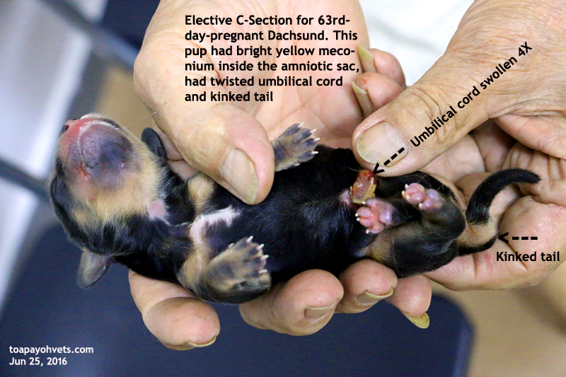
4 days have passed. What happened to the puppy with the crooked tail and swollen umbilical cord?
This morning, I visited the C-section 634rd-day pregnant Dachshund. She's OK but was not permitted to nurse her own new born puppies. The poodle was the surrogate for the 3 puppies. The 4th with the crooked tail and swollen umbilicus had passed away the 2nd day although the umbilicus had dried up.
"I had tied up the intestine which was inside the swollen umbilical cord," the breeder said. "So the intestine was deprived of the blood supply, killing the puppy. I had a few of such cases."
Each breeder or vet has his or her own theories about medical conditions and so I did not say that this puppy had been severely stressed as the amniotic sac was filled with yellow meconium unlike the normal 3 siblings. In human medicine, ultrasound scans showing meconium in the baby will lead to an emergency Caesarean section to save the baby. But costs prohibit the breeder from spending on such ultrasounds.
The umbilical cord was twisted and so it swelled 4X. But I know the experienced breeder would not accept my theory and so I did not expound my theory. "Maybe, next time, I should check the swollen umbilical cord for any intestines inside and push them inwards before you tie the cord," I said.
"I should assist you during C-sections," he said as I usually asked him to be outside the operating room unlike one lady vet who had 100% no-death in puppies in C-sections with him and so she was an excellent vet. Unfortunately she had retired from doing C-sections for him and he had been to various vets throughout the past years. He advocated an assistant to elevate the lower body of the dam so that C-sections can be completed faster. Or the assistant holding part of the uterine horn while I take out the puppies faster.
The latter suggestion was preposterous as the gravid uterine horn is not that long unlike that of the cow's. There is just no space for another assistant to hold part of the uterus while the vet milk out the puppy.
Many sensitive younger vets may not be happy with his presence or suggestions of assistance or his criticism of puppy death being due to delay in pulling out the puppy and so the puppy had an overdose of the isoflurane and therefore die the next day. I don't permit outsiders into the op room during C-sections to keep the room as sterile as possible.
In this 63rd-day pregnant, the breeder had outsourced the 3 puppies to the poodle and is pleased with his decision as the puppies are thriving. He believed that the other Dachshund had toxic milk killing all the 3 puppies I delivered alive on C-section and so was not taking chances with this Dachshund. This Dachshund had snow-white milk whereas the other had no milk and the next day, produced thick yellowish milk deemed toxic to the 3 puppies.
Breeders have their beliefs. He even suggested that a longer incision bringing out the whole gravid uterus would compromise the milk production of the mammary gland and so vets who incise the abdomen from umbilicus to pelvis as stated in the vet books have had damaged the milk system, leading to poor quality milk production.