Pet health and care advices for pet owners and vet students, photography tips, travel stories, advices for young people
Tuesday, September 5, 2017
3132. Ear abscesses in hamsters - cases at Toa Payoh Vets
Hamsters do suffer from ear warts and ear abscesses but few owners know. Below are some images of cases.
1. For ear warts, the only treatment is surgery. No medication will make the wart disappear.
However, some vets may prescribe medication as they do not perform surgery.
--------------------------------------------------------------------------------------------------------------------------
Monday, September 4, 2017
3132. A guinea pig had watery greyish stools for one week.
Histology shows benign haemangioma. The guinea pig went home after surgery 2 weeks ago. He had poor appetite in the first week. Stools were normal. The owner fed other food like vegetables. Also spray anti-mange and mite from a pet shop, onto the body filled with patches of hair loss. The guinea pig must have licked the spray causing gastroenteritis. He eats the watery stools as well, causing more watery diarrhoea.
Hospitalised one day. Stools back to normal as shown. Guinea pigs do eat their stool pellets to redigest the pellets, as part of their nature. Now the stool pellets are normal and so it is OK for him to eat them.
Saturday, September 2, 2017
Tuesday, August 29, 2017
3128. CASE STUDY. VIDEO SCRIPT. A Syrian Hamster has 2 large tumours below the throat
"I note that you can remove a hamster's tumour in 5 minutes," the young lady had decided not to let the first vet operate on her Syrian hamster with 2 large subcutaneous tumours below the neck. The hamster had been scratching the affected neck area bald but her appetite is excellent.
If the tumour can be removed in 5 minutes as contrasted to an hour's operation estimated by the first vet, that means that her hamster would likely not die on the operating table. The owner had to sign a consent form for anaesthesia and surgery and she was aware of the anaesthetic death risks.
"5 minutes is possible if the tumour is as small as 5 mm across, but your hamster's tumours are over 2 cm across," I said. "It takes much longer than 5 minutes."
The first vet had warned her: "The tumours are near the major blood vessels and nerves of the neck and the hamster may die during excision of the tumours!"
"What the first vet says is correct," I replied. "If you had got the tumours excised by the vet when it is very small, less than 5 mm across, the risk of damaging the blood vessels and nerves are practically nil."
INTRODUCTION
Hamster tumours usually develop as small lumps in hamsters over one year old. Many young Singapore hamster owners live hectic working and personal lifestyles. Some do not even have time for their retiree parents, let alone the pet at home.
Singapore has so many distractions and entertainment outlets for the young hamster owner and so the hamster's tumour is left to grow.
VIDEO OF SUNTEC CITY
Many young Singaporeans procrastinate visiting the vet till the tumours are gigantic as in this case and many others seen at Toa Payoh Vets.
(10 images of hamster tumours operated by Toa Payoh Vets over the 10 years). Many vets do not wish to operate on large hamster tumours. Some will advise oral medication and euthanasia when the infected ulcerated tumours are too large causing the hamster poor health.
Surgical excision is the only treatment. Some vets don't perform hamster surgeyr.
DIAGNOSIS AND TREATMENT by electrosurgery
This Syrian hamster had 2 mobile subcutaneous tumours. The smaller one of 1-cm across is nearer to the major blood vessel. The larger one of 2.5 cm across is below the throat. This is not going to be a simple operation as the owner expected the hamster to be alive at the end of the surgery.
Surgical excision is the only option. Zoletil 100 IM is given at 0.01 ml and it was sufficient for me to remove the 2 tumours without the need for topping up with isoflurane gas. I undermine the skin to detach the tumours. Then I clamp the base and excised the tumour using electro-excision to clot the area. For the larger tumour, I ligate the stump of the large tumour to prevent any bleeding. There was no need to ligate the stump of the smaller tumour. There was no bleeding. 3/0 absorbable sutures were used. The skin was closed by continuous sutures.
VIDEO FOOTAGE
POST-OPERATION
The Syrian hamster was all right and went home around one hour later. The owner and her Taiwanese mother were pleased and would give the oral antibiotics and painkillers. It is a joy to see the Syrian hamster alive.
CONCLUSION
Anaesthetic death is possible if large tumours are operated as they take a much longer time.
Do not procrastinate as hamster tumours never disappear but will grow larger daily.
A 5-minute surgery is possible if the tumour is less than 5 mm across and the risk of anaesthetic death is practically zero. In this case, the hamster survived the anaesthesia and lived to an old age.
.
------------------------------------------------------------------------------------
OTHER IMAGES NOT FOR THIS VIDEO
Thursday, August 24, 2017
3127. INTERN. Single pup syndrome - Expensive Dachshund puppy saved by Caesarean section
E:\2017TPV_JUL_SEP\20170718Birthday_LP
July 18, 2017 Elective Caesarean Section. Judgment of breeder to save the pup as it is too large to be born naturally.
2064 Breeder worried. No strong uterine contractions. One large pup palpated. Single pup syndrome. At Toa Payoh Vets now.
2065. Prepare for surgery.
2070 Cone mask. No IV sedatives like propofol. Just isoflurane and oxygen gas only. By cone mask first, then intubate with endotracheal tube and 2-2.5% isoflurane gas for maintenance.
2071. Intubated.
2072. Uterus taken out of the body.
2073. Stiching in 2 rows. Illustrations shared with the Corgi pup C-section
2074. 2nd row completed.
2075. Dam awake fast. Washed. oxytocin, antibiotic and painkiller injections.
2076. Footage of normal water bag with clear fluid inside. Placenta is not normal firm and turgid in size but "melting". The C-section was just in time.
2083. single pup alive.

FOLLOW UP 3 WEEKS LATER visit to breeder's farm
See footage in Corgi video in the other blog.
FOLLOW UP AT BREEDER'S FARM 3 WEEKS LATER.
PUPS OK.
E:\2017TPV_JUL_SEP\20170806Review_3_C_sections
2750 Edit 00:00 - 1:26
Footage shows one of 3 Corgis thriving
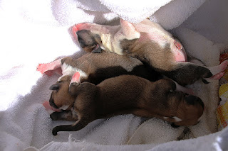
Footage shows the single pup (Dachshund) thriving under the poodle. Its mother has not sufficient milk.
The poodle is the surrogate mother of this single pup syndrome Dachshund and one Corgi. The other 2 corgis are nursed by the Corgi dam.
2773 2 Corgi pups
2772 Dachsund single pup syndrome
------------------------------------------------------------------------
CONCLUSION
LIFE AND DEATH depend on the judgment and knowledge of the breeder.
Some do wait for the pup to be born naturally. Generally, the single pup syndrome requires an elective Caesarean section as it is too big to be born naturally in small breed dogs. Death of the pup is expected if the breeder waits past 70 days. The suitable time to operate is 63 days when the pup is fully formed. The general time of birth for dogs is 59-63 days.
.

3126. INTERN. Unusual case of one uterine horn filled with dead pups, one with normal pups
The Corgi's uterus consists of 2 uterine horns. The left and right uterine horns.
(Image). Normally both uterine horns will be filled with healthy pups in clear water sacs.
In this unusual case,
The entire left uterine horn was filled with black fluid and 3 dead pups and melted placentas.
The entire right uterine horn was filled with normal pups in clear water sacs and placenta.
E:\2017TPV_JUL_SEP\20170716Sun_C_section_mummified_L_horn
Videos
2011 Black vaginal discharge. Breeder phoned Toa Payoh Vets for an emergency Caesarean section.
2012 + 2013 Prepare for surgery. IV drip, isoflurane gas anaesthesia only. No sedatives like propofol IV.
*2014 Left uterine horn. Black fluid filled the inside of the uterus. All 4 pups dead, mummified with no placentas.
Image
Right uterine horn
2016. Right uterine horn is ready for incision
2017, 2018 normal pups pulled out.
Illustration of stitching showing 2 rows
*2019. Uterine horn stitched up in 2 rows of inverting stitches.
lst row is parallel to the incision
*2020 lst row completed. Footage shows 2nd row started.
*2021 end of stitching of 2nd row
2022 End of surgery. Dam cleaned up. Oxytocin, antibiotic and painkiller injections will be given.
2023 + 2024 Healthy pups from right uterine horn.
2029, 2034 Goes back to breeder farm
FOLLOW UP AT BREEDER'S FARM 3 WEEKS LATER.
PUPS OK.
E:\2017TPV_JUL_SEP\20170806Review_3_C_sections
2750 Edit 00:00 - 1:26
Footage shows one of 3 Corgis thriving
Footage shows the single pup (Dachshund) thriving under the poodle. Its mother has not sufficient milk.
The poodle is the surrogate mother of this single pup syndrome Dachshund and one Corgi. The other 2 corgis are nursed by the Corgi dam.
2773 2 Corgi pups
2772 Dachsund single pup syndrome
Wednesday, August 23, 2017
3126. Intern Images*****. Be Kind To Pets Veterinary Educational Images - dogs,cats, hamsters, people
https://2010vets.blogspot.com/2018/09/3282-interns-be-kind-to-pets-images-sep.html
DOGS
HAMSTERS
LIZARDS
TURTLES
PEOPLE
https://2010vets.blogspot.com/2018/09/3282-interns-be-kind-to-pets-images-sep.html


