After 7 days of antibiotic and painkiller treatment as an in-patient, this rescued young Persian's damaged infected eyeball was removed by Dr Daniel.


| TOA
PAYOH VETS toapayohvets.com Blk 1002, Toa Payoh Lor 8, 01-1477, Singapore 319074 Tel: 6254-3326, 9668-6469, 9668-6468. judy@toapayohvets.com 22 May, 2015 Focus: Small animals - dogs, cats, guinea pigs, hamsters, turtles & rabbits |
|||||||
|
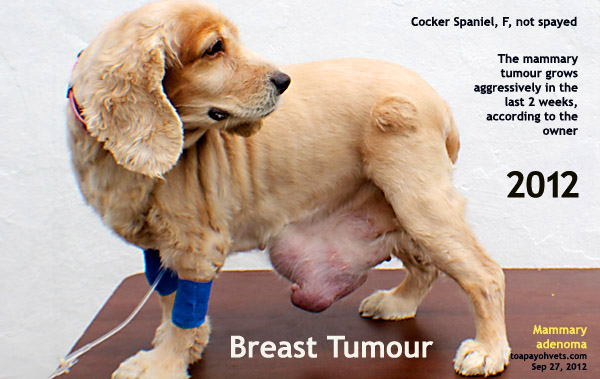
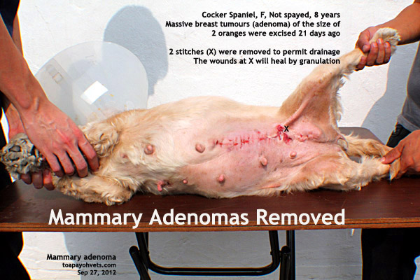
Mammary adenomas in an American Cocker Spaniel Dr Sing Kong Yuen, BVMS (Glasgow), MRCVS First written: 21 September, 2012 Update: 22 May, 2015 "Why did you delay seeking surgery till the breast lump is gigantic in size, like two tennis balls combined?" I asked the Buddhist temple teacher who taught Buddhism and morals to over 800 children of devotees. "The breast lumps grow very fast in the last 2 weeks," she replied. "Nobody in the family wants to bring the dog for surgery. My brother said to let her die." The solution was surgical removal but the family did not want to pay the medical costs. She was kind to enough to bring in this old dog for surgery at Toa Payoh Vets.
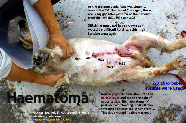
I tacked in "walk-in sutures" through the muscle layer and then the skin, using 2/0 sutures to ensure that the large long wound would not break down.. The dog came back 4 days later. There was a swelling at one end of the tumour. " "There's seepage at the left Mammary Gland 5 area," I said to Dr Daniel who is in charge of the case. "Just cut off two stitches," I said to him. "In this way, the serum can drain out. No need to re-stitch." After that, we had no more visit from the Buddhist teacher. I was glad that this dog was not abandoned when she had the tumours. Such incidents do occur in Singapore. It is best to spay the female dog when she is young as breast tumours are less likely to develop in spayed dogs.
More case studies, go to: Cats or Dogs Make an appointment with your vet. Or tel 6254-3326, 9668-6469 for an appointment to discuss health screening for your senior companion. Or e-mail judy@toapayohvets.com your requirements. |
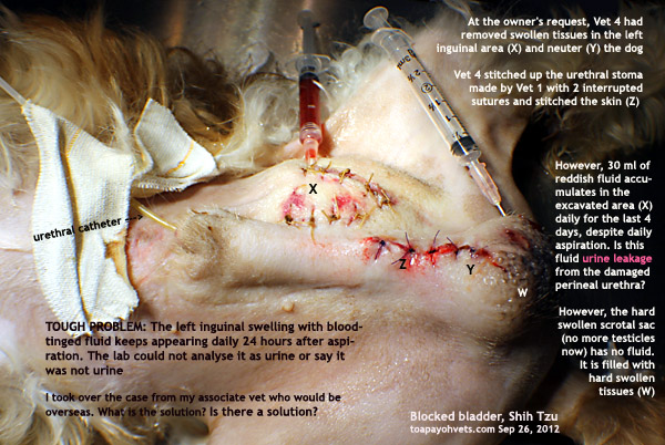
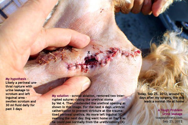
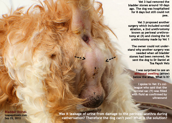 This is one supporting evidence and there must be others to substantiate this urine leakage (ultrasound of scrotum filled with fluid), swelling of left inguinal area near where Vet 3 made a skin incision to access the bladder for urinary stone removal daily with blood-tinged fluid while the dog was catherised with a smaller sized urinary catheter
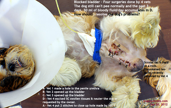 | 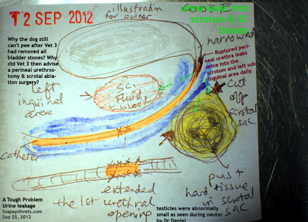 |
| Large left inguinal swelling due to urine + blood accumulation | Problem and surgical solution explained by illustration for the owner |
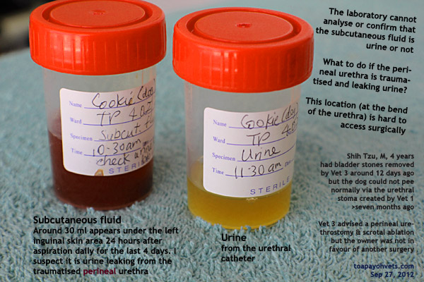 | 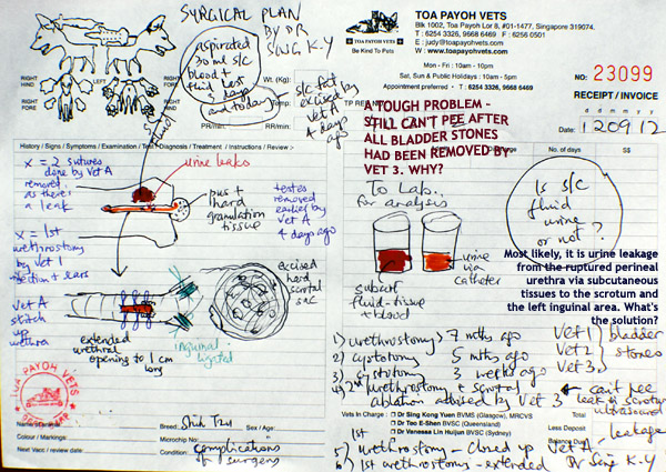 |
| Inguinal fluid and urine for lab analysis | Surgical plan from Dr Sing Kong Yuen |
| The dog still had difficulty in urination after Vet 3's surgery. My surgical approach to post-op complications from Vet 3's surgery which had removed the bladder stones | |
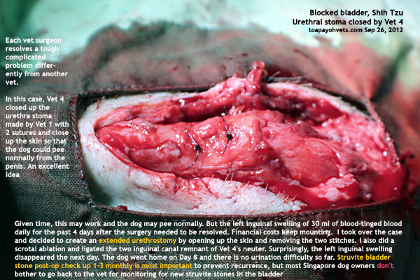 |  |
| Open up the wound | Scrotal ablation |
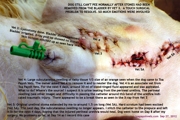 |  |
| Enlarged Vet 1's urethrotomy incision | Ensure urethra is patent |
 |  |
| Healing of the surrounding areas | Goes home with prescription diet S/D |
 | 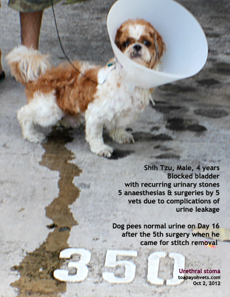 |
| Dog is able to pee via the original enlarged urethrostomy for the past 3 months and as at Jan 7, 2013. Dog eats prescription diet C/D. Urinary analysis and X-rays on a regular basis are recommended. Will the owner comply? | |