Monday, October 24, 2011

703. An old dog with a breast tumour bigger than 2 golf balls

"I went to Vet 1 as my dog was bleeding," the lady was concerned about the bleeding in her 12-year-old Shih Tzu rather than the tumours. "The vet bandaged the lump."

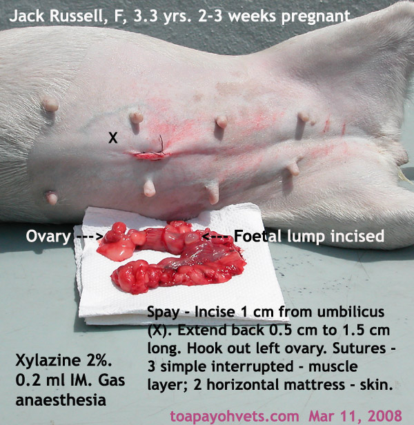
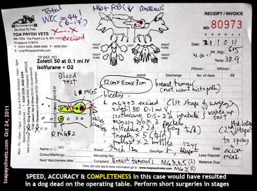
"Your dog has at least 4 breast tumours," I said. "How long did the biggest tumour take to grow?"

"Very fast," she said. "In the last month, it just grows so big." The tumour was the size of two golf balls. This was the left MG5 (the left caudal mammary gland, 5th gland)

HOW THIS CASE WAS HANDLED BY ME & DR VANESSA

1. Blood test compulsory.
Total white cell count was over 42 indicating a bacterial infection. I had the dog on IV drip and baytril overnight. Some owners object to blood tests due to financial considerations. X-ray of the chest would be preferred too but this is often not asked of the owner so as to reduce costs.

2. Surgery
Operation to be done the next day due to economic reasons. I prefer wait one week on antibiotics but there are such cases where the owner does not want to wait. Sometimes it may be better to reject such cases but compassion and the interest of the sick dog must be considered by the operating vet.


3. Pre-surgery - Zoletil
No sedation preferred. Just use isoflurane gas + oxygen.
Zoletil 0.1 ml IV although Dr Vanessa would have used Domitor. Isoflurane gas + oxygen via mask and then intubation. "Zoletil is safer in old dogs," I said. "Few vets I know use it for small animal sedation."

4. Short surgery
Only the big tumour was excised. This already took around 45 minutes. If the vet attempted to excise all, a dead dog would be the result as old dogs are very high anaesthetic risks.

In a recent on-going court case of a death of a man in his 40s who had liposuction and fat transfer to his chest, the general practitioner (GP) taking 3.5 hours for lipo-suction and transfer of fat to the chest of a man who died, I believe that the whole process of IV anaesthesia (involving topping up) took too long and lead to death. However, there were numerous perforations of the internal organs.

From what I read:
a) The expert witness mentioned about over-sedation as a cause of death as the patient did not respond even though he had numerous internal punctures.
b) The GP doctor doing the liposuction said death was due to fat embolism.
c) The GP doctor doing the IV anaesthesia (Propofol sedation given to effect iv and oxygen, but no general anaesthesia) said that the patient was alive at the end of surgery but died around half an hour later. Therefore anaesthesia was not the cause of death.
d) The autopsy report said death was due to numerous perforations of the intestines and the liver.
e) The judge was exasperated as to why the time and dosages of Propofol had not been made available before the inquest. The info was said to be in the GP's hard disk during the inquest. The case is on-going.

5. Anaesthesia
I was in charge of anaesthesia. The maintenance dose was 0.5 to 1.5% isoflurane. Towards the end of 30 minutes, I showed my assistant Min that the tongue has become white at the edges despite just 1.5%. I reduced to 0.5%. Switched off at the 2nd last stitch. The dog moved a bit. But stitching was done and the dog woke up. This was good anaesthesia. No delay in waking up and no need to use Antisedan as in the case of domitor.

6. Speed of surgery
6.1 Know your anatomy. There is the big blood vessel at around MG3 and MG4. Slow dissection and undermining is OK if you have time but old dog surgery need to be completed in less than 30 minutes for good anaesthetic outcome.

6.2 "No point having a perfect stitching and surgery if the dog dies on the operating table," I said to Dr Vanessa who started stitching up from one end intending to stitch at 0.5 cm intervals as this was her usual method. The wound was around 20 cm long. "Stitch at 1 cm intervals." I also advised stitching at the major points first e.g place 4 stitches and then continue stitching in between.

6.3 "Use walk-in sutures," I explained. But Dr Vanessa was comfortable in using subcuticular sutures to hold the subcutaneous fat together. This took much more time as skin sutures were then sutured with simple interrupted sutures. In "walk-in sutures," I would stitch the skin, then subcutaneous fat and then skin all in one movement using horizontal mattress sutures on the skin.

OUTCOME
The whole process was completed by Dr Vanessa in around 45 minutes. Any longer would have caused death, in my opinion as the tongue was turning pale pinkish cyantotic and white at the edges.

The dog was alive and that was what the owner expected. She had come in the morning of the surgery on Day 2 to ask more questions. I had to be frank as I told her that I needed time to prepare for surgery.

In the evening the happy owner took the dog home. I write this case as a mentoring case done by me.

QUESTIONS ASKED
1. The financial considerations which were addressed yesterday.
2. The chances of survival. Hard to predict as the dog had a bacterial infection and was old at 12 years. Short surgeries make survival likely. So, the other tumours and spaying of the dog had been advised for two weeks later.
3. Whether the breast tumour is cancerous. Most likely as it grows explosively big within a month.
4. Will the tumour come back? Most likely as it is cancerous. However, the owner does not want histopathology by the lab to confirm it.


CONCLUSION
No matter how great the vet surgeon is, anaesthesia is the most important in any surgery as a dog alive is what the owner wants. Beautiful surgery is a bonus.


Toa Payoh Vets webpage is at:
http://www.sinpets.com/F6/20111035old-dog-zoletil-breast-tumours-singapore-toapayohvets.htm

Sunday, October 23, 2011

Keep Spay Surgery Simple - No subcuticular suturing

I am writing to share my over 30 years of experiences in spaying dogs and cats done by me and by other vets in Toa Payoh Vets.

I started Toa Payoh Vets in 1982 and it is 2011 now. I have seen a few cases of stitch breakdown using the method as advised by the university professors and veterinary surgery text books (Method 1).

The vet university professors since my time in 1974 when I graduated from Glasgow University and even recently in Australian Universities, advocate the following steps after removing the ovaries and uterus:

1. suture the linea alba
2. suture the subcutaneous fat to seal up dead spaces
3. suture the skin
The theory is sound and many newly graduated vets adopt this system (Method 1). Over the years, I encountered a few cases of stitch breakdown and infection in dogs and cats using Method 1 done by other vets. Or intense redness and inflammation at the surgical area.

My method does NOT use Step 2 and I have less than 1% of post-op stitch breakdown. I have no figures to substantiate my claim. I am merely sharing my experiences as I am in my retirement era, being 61 years of age.

I just hope that this knowledge may benefit the dogs and cats. In the early years of 1970 - 1980, I did not prescribe pain-killers unlike nowadays and I don't have many post-op spay problems. Times have changed. Pain-killers inclusive of antibiotics are a must.

There are many reasons for stitch breakdown but one of them is the irritating subcutaneous suture. The dog or cat keeps licking the wound or just use the hind paws to scratch it. E-collars do not help much. Soon, the unhappy owner brings in the dog or cat for review.

In private practice, the vet earns his reputation by not getting less or no post-op spay stitch breakdown as owners will compare and complain. Obviously, in other surgery like breast tumour removal, subcuticular sutures are needed to close the dead spaces. I use "walk-in" sutures. This involves suture needle going into the skin, then into the subcutaneous fat and out of the subcutaneous fat, then out of the skin on the other side. From there, I transverse, needle goes into the skin, subcutaneous fat and out from the initial side. This makes for a horizontal mattress skin suture as seen from the skin. In this method, surgical time is reduced, unlike separate subcuticular sutures of the subcutaneous fat. See case report at:
http://www.bekindtopets.com/dogs/20081235Mastectomy_Female_Dog_ToaPayohVets.htm

Then simple interrupted sutures of the skin as this is a usual method by many vets.

The two methods of SPAY IN THE DOG AND CAT are illustrated by real cases as follows:

METHOD 1 - BY THE (VETERINARY SURGERY) BOOK


1298. Subcuticular sutures have been used. The stitches under the skin hinder healing due to infections or traumatic injury from licking.
1299. Stitch breakdown and infection. Re-stitch again but no more subcuticular suturing.
METHOD 2 - BY DR SING


1296. A spay hook enables the vet to make spay incision small in the dog and cat.
1297. Dr Sing's dog spay procedures. No subcuticular sutures.
All the years, I use absorbable PDS sutures and this means the owner does NOT need to return for stitch removal. I just use one packet of sutures. However, with Method 1, I note that some of the younger vets must use two packets and sometimes three packets in dogs that are pregnant and of bigger breeds.

I have seen cases where a vet uses 3 packets of sutures to spay a medium sized dog. This is definitely too many. One packet is chromic catgut. Two packets of absorbable monofilament. With my method, one packet of absorbable suture is sufficient for dogs up to medium size, using the spay hook.

Keep spay simple. I hope this report is of use to some practising vets. It cuts down on expenses in using more sutures and in private practice, costs keep rising as bureaucrats think of ways and means to generate income from the industry by increasing regulatory fees. Keeping spay simple and using walk-in sutures make economic sense as well as keep owners of dogs and cats happier.

OTHER INFORMATION


4448. KPI - How long it takes to spay a female dog? 26 or 18 minutes?
4451. Surgery Audit: How long it takes to spay a female dog? 18 minutes


4438 - 4444. When do you remove the dressing or stitches after spay/neuter?
4475. Never discharge a spayed dog with a hole in the operation area
 
Toa Payoh Vets webpage is at:
http://www.sinpets.com/F6/20111033spay-cat-dog-no-subcuticular-singapore-toapayohvets.htm

701. Follow up on: Pterygium + Glaucoma + Haematoma in 2010, Enucleation in 2011

What is a pterygium? It is an invasive proliferative fibrovascular growth.
Excision of the pterygia was requested by the owner as the dog had difficulty seeing in the right eye and kept rubbing the right eye.

Excision was by electro-surgery under general anaesthesia. In human medicine, there are various sophisticated surgical methods used

I find that many old dogs can survive if the surgery and anaesthesia can be done under 15 minutes. Provided there is care in the anaesthesia and knowledge of emergency resuscitation. The anaesthetic time would be prolonged as there were two pterygia to be excised.

The Divine Powers above threw in an ear haematoma to be operated too. That meant a much longer anaesthetic time.

The longer the anaesthesia, the higher the chances of the old dog dying on the operating table.

This dog needed over 60 minutes of surgery. Would his cornea rupture? That would increase the anaesthetic time.

The dog did not wake up within 2 minutes at the end of anaesthesia.

An antidote (Antisedan) woke him up. He recovered smoothly on Domitor pre-anaesthetic dose, isoflurane gas maintenance and Antisedan anti-dote. He woke up quietly as if he had a good nap.

Fortunately, the mission was accomplished with excellent anaesthesia. This story had a happy ending for everyone. Further info is as follows:

In March 20, 2003, I saw a Shih Tzu puppy with a growth on the right eye for the first time. Was this a rare case of a pterygium? No. It was a Dermoid as it had hairs and was of a different colour. See: Mar 20 03. The Shih Tzu puppy has a "pterygium"?
7 years had passed since I saw the afore-mentioned Shih Tzu puppy with the Dermoid. A real case of pterygium surfaced in February 2010.

She was more knowledgeable than me in specific area affecting this very gentle X-bred dog, as she correctly stated: "My dog has a pterygium," when she first consulted me.  She requested me to excise the two bulging growths on the cornea of her dog.

The dog also had glaucoma. She was old. There was a big risk that the cornea might rupture. The bigger risk was death on the operating table as this old dog would be a high anaesthetic risk. Every old dog under anaesthesia is a big risk and deaths from anaesthesia are very unpleasant for the vet as the whole family never forgets nor forgives when the outcome is a dead body. I was very apprehensive that this well cared for and loved family member would die on the operating table as anaesthesia would take a long time to excise two pterygia. As if my worries were not sufficient, the dog also had a right ear haematoma to be operated upon. That meant that the surgery would take over an hour!

The longer the operation, the higher the chances of deaths on the operating table.

"Why do you want the pterygia removed?" I asked the determined owner.
 "My dog walked towards her left when the pterygia started to grow fast towards the centre of the cornea. She cannot see from her right eye as the pterygium had obstructed her vision." the owner said. "In addition, she keeps scratching her right eye."

"If the cornea ruptures during excision, I will have to remove the whole eyeball," I said. "Yes," the lady consented. There was this understanding and awareness of the high risk of the cornea bursting when I excise the pterygium when the intraocular pressure was very high. One pterygium was already worrisome. But this dog had two big ones.

"Have you considered enucleation (eyeball removal)?" I asked as glaucoma can be a very painful condition and treatment using eye drops must be given regularly and drug control may become ineffective over time. I don't know much about its glaucoma formation as it was treated by another vet. Glaucoma was controlled by eye drops.

"The other vet has recommended it," she said. "But my family does not want the dog to lose his eye." So enucleation was out of question.

As to what caused such a rapid growth of the pterygia in this beloved dog, it is hard to say. In people, pterygium is said to be caused by ultra-violet light exposure.

It was fortunate that the old dog survived the anaesthesia and went home. "Take good care of her glaucoma," I said to the happy lady as I discovered that the old e-collar was no more worn. I had asked whether she still had the e-collar and she said it was worn out and disposed. "That means that she must wear the e-collar all the time for many weeks and stay out of the sunlight and wind." 7 days later, I would need to review this case.

Pterygia Surgery at Toa Payoh Vets


Haematoma Surgery At Toa Payoh Vets


Toa Payoh Vets webpage and images for this article is:
http://www.sinpets.com/F6/20111023pterygia-glaucoma-haematoma-dog-toapayohvets-singapore.htm

FOLLOW UP IN 2011
In 2011, the dog's eye was enucleated. The webpage at Toa Payoh Vets is at:
http://www.sinpets.com/F5/20111031old-dog-pterygium-glaucoma-anaesthetic-reactions-success-singapore-toapayohvets.htm

Friday, October 21, 2011

700. Follow up on paper training and stool eating puppy

LATEST E-MAIL REPLY FROM DR SING DATED OCT 21, 2011

Paper shredding is a common complaint in paper-training. Taping the edges is one method which you have used successfully.

Stool eating is a difficult problem to solve as the cause is usually unknown.
1. Pepper, chilli sauce and others have been added onto the stools without effect.
2. Some owners buy the "anti-stool" eating powder from the pet shops and claim effectiveness as long as the powder is eaten.
3. Others add pineapple slices onto the food and claim good results.
4. Distraction methods: Another writer advise putting a can of coins on top of the door. Hide somewhere. When the puppy eats the stools, the can of coins drop onto the floor. Personally I doubt that the puppy (as a canine with sensitive hearing) will not know that the owner is hiding somewhere.
5. Another method is to squirt water onto the puppy when he eats his stools. You need a water gun.
6. As you can see, it is extremely difficult to resolve the problem. Picking up the stools immediately (without making a fuss) seem to be another method. In this way, it is hoped that the puppy outgrows this bad habit. Otherwise he continues the habit to old age. This appears to be what you are doing.
7. My idea is to take the puppy outdoors to poop after meals but this may not be practical for you if you live in a high rise. This involves a routine as follows: Puppy completed eating. Take him outdoors to the grass. In short, take the puppy to another area (e.g. balcony has newspapers with smell of poop) to poop. This works if the puppy poops within 10 minutes after eating and the owner is free to do this consistently for 2-4 weeks.


----------------------------------------------------------------------------
On Thu, Oct 20, 2011 at 9:50 PM, A@gmail.com> wrote:

Hi Dr Sing,

I hope this email finds you well.

It has been almost a week since we tried your suggestion. Newspapers (full page, spread out) are placed on the top right corner of the toilet and the puppy eliminates at the area. We are slowly trying to reduce the area of the newspaper. Attached is a picture of the toilet for your reference.

However, he has started to (in this order, not sure if it tells you anything) smell, lick and eat his stools. Soiled papers are changed almost immediately in the day, as someone is usually around to hear him running on the newspapers. One of us will wake up in the middle of the night every few hours to check on him and clear any waste, but sometimes he has already finished eating his stools. We are cautious not to disturb him when he eliminates. Pepper does not help as he eats his stools which has pepper. Is there anything we can do about this?

As the puppy loves to shred newspapers, we use masking tape and lace it white vinegar to prevent him from peeling off the tape. It works most of the time, and he does his business as usual, including eating his poop.

Once again, thanks for your advise and I hope to hear from you soon.

Best regards,
A
------------------------------------------------------------------
From: A
Sent: Friday, October 14, 2011 10:00 PM
To: Kong Yuen Sing
Subject: Re: paper training problems for a 3 month old poodle

Hi Dr Sing,

Appreciate your prompt reply, we are currently trying it out and will let you know how it goes. Have a nice weekend. :)

Best regards,
A

--------------------------------------------------------------------------

From: Kong Yuen Sing
Sent: Friday, October 14, 2011 7:04 AM
To: A
Subject: Re: paper training problems for a 3 month old poodle

Thank you for your detailed e-mail and the 3 images. As every puppy and owner is different in personality and training experience, the following reply may or may not be useful to you.

1. You have confused the puppy in attempting to grate-train (using the cage) and paper-train him (inside the at the same time. Puppies, like babies, are creatures of routine. One method of toilet training is needed. Either grate-training or paper training.

2. At the pet shop, the puppy has had been trained to eliminate in the grate (floor of the crate or cage). This type of grate+pee pan housing is prevalent in the Singapore pet shops as shown in the image you posted to me. The puppy wanted to eliminate on the grate but you wanted him to do on the paper and most likely paid extra attention and fuss. So, the puppy was not free to eliminate in privacy after eating as in the pet shop.

3. To make a long story short, I would propose just write about paper training as I presume this is what you want to achieve instead of writing about grate+pee pan training or both methods. The grate+pee pan method involves buying the grate + pee pan separately or the use of your existing crate.

PAPER TRAINING

3.1 You confine the puppy inside the toilet. Install a baby-gate. Cover the toilet floor 100% with paper. At one corner (e.g. left half, back), away from the baby gate, put a piece of paper with the puppy's urine or tissue paper with his urine smell. At the other corner (e.g. right half, front), his food and water. In the right half, place his towel, bed, or floor mat, for example. This will be his clean area.

3.2 The puppy is left there for the next 2 - 4 weeks and given privacy, not stressed out by your scoldings or presence or the presence of children and family members during eating for at least 1 hour.

3.3 The puppy can be taken out to play after he has pooped regularly and this may take a week. This is where you observe signs of elimination (squatting, turning) and put him quickly back to his toilet.

3.4 Change soiled papers frequently.
3.5 Within 7 days, you should see that the papered corner where the puppy pees and poops will be. Reduce the paper area.
3.6 If the puppy shreds the paper, you still need to persevere and do not scold or shout at him.

In conclusion, confinement for the first 2-4 weeks is the key to success in toilet training. What you do during the confinement will require your perseverance, hard work of keeping the puppy's "den" (toilet) clean.

The training is much more than what I write. The devil is in the execution. Pl let me know when you have succeeded or if you have more queries.

Best wishes.


-------------------------------------------------------------------------------


On Thu, Oct 13, 2011 at 9:34 PM, .........@gmail.com> wrote:

Dear Dr Sing,

I would like to seek your advise with regards to paper training. Three pictures are attached for your reference; a picture of the cage he sleeps in, the toilet area with newspaper, and a view of his cage from the toilet.

I have a 3 month old male miniature poodle whom I brought home about a week ago from a pet shop. We bought him a cage and are in the process of training him to eliminate in the toilet on the newspaper. At the pet shop, he would eliminate about 15 minutes after his meal. However, he eliminates only after a few hours. For example, we will give him dinner at 7.30PM and most of the time he will not eliminate until the next morning. It is rare that he will eliminate within 2 hours. When we spot signs that he wants to eliminate when he is outside the cage, we will bring him to the toilet and coax him to eliminate, whether big or small ‘business’. Most of the time he will not do it unless he has not gone into his cage for a very long time, say a few hours. There are times when he will run to the toilet and smell the newspapers, only to run out, which we take as a hint that he wants to eliminate and put him into the toilet telling him to pee, using the same word every time. Rewards given when he does the right thing.

When we put him into the cage, he will eliminate within a few minutes or even seconds when we are not looking, and that is after we have just brought him to the toilet. The areas which he eliminates in the cage are not consistent. Even if he does not intend to eliminate, he will turn in circles and give us false alarms, as I believe he knows we will bring him out when he does that. We tried washing his entire cage with antiseptic but he still does it inside the cage. The newspapers in the toilet have traces of his pee and also a kind of liquid we bought from the pet shop to help him recognise the place.

For the first two or three days, he used to pee all over the place but after scolding him a few times and cleaning with vinegar, he has not done it since. A crate didn’t help as he peed inside it.

Are we doing something wrong? Some expressed their opinion that a pee tray would be better and the distance between the toilet and cage is too far for him, making him confused. He has not had his third vaccination so we have yet to bring him out.

Thank you very much for taking time to read this email, and also for your informative articles. Looking forward to hear from you soon!


Best regards,
Name


DETAILS AT TOA PAYOH VETS
http://www.sinpets.com/F5/20111023puppy-toilet-training-paper-or-grate-not-both-singapore-toapayohvets.htm

Thursday, October 20, 2011

699. Liposuction Death - Practice guidelines for safe sedation

Straits Times, oct 20, 2011 Pg A4
Liposuction Death - I did not cause death, says doctor

Patient: Mr Heng went to Reves Clinic in Orchard Rd on Dec 30, 2009 for a liposuction and fat-transfer operation.

Doctor 1: Dr Jim Wong did the surgery. Set up nfusion pump which administers drugs including propofol into the patient.

Doctor 2: Dr Zhu Xiu Chun @ Dr Myint Myint Kyi - monitor the patient's vital signs. Continuous oxygen supply, equipment measuring the vital signs was in place.

Said readings were within an acceptable range, no snoring or other abnormal respiratory movement to sugestg that Mr Heng' airway was disturbed. If the patient did not move much, he was quite sedated and dosage of propofol would be adjusted lower. If he moved or made sounds, after making sure that vital signs were staable, the dosage would be increased. Moderate sedation - responsive to tactile sensation.

Procedure: 12.30 pm to 3.50 pm
25 minutes after end of procedue, Dr Zhu was told there was an emergency. Mr Heng was pale and unresponsive and died.

Autopsy: >10 puncture wounds in intestines, liver

Question: Mr Heng's rep said that Mr Heng did not show any overt signs of pain because he was more than moderately sedated as multiple punctures in his internal organs from liposuction would be very painful.

MINISTRY OF HEALTH GUIDELINES FOR SAFE SEDATION

1. The changes in dosage of propofol had NOT been recorded. This was unsatisfactory.
2. The patient's vital signs were recorded every 15 minutes. Every 5 minutes would be better. Vital signs include pulse rate, blood pressure, oxygen saturation

In an earlier inquest - fact finding rather than determination of who was to blame for Mr Heng's death, two experts mentioned that the patient was over-sedated.

698. Pterygia and glaucoma - anaesthesia in a 12-year-old dog

In Feb 2010, I operated on the old dog with two pterygia in the right eye.


The case is described at:
http://www.asiahomes.com/singaporetpvet/rabbit/rabbit.htm


Yesterday, Oct 19, 2011, I operated on this 12-year-old again. She was a happy dog as he wagged her short tail, unlike some dogs that had to be dragged into the vet surgery.

The pterygia had re-grown and covered the whole eye. Despite anti-glaucoma eye drops given by the medically knowledgeable owner, the glaucoma could not be cured. Now the ptergygia had covered the whole cornea as well, nearly 100%.

"I thought you spoke to the human eye expert regarding opening a hole in the sclera to drain the high eye pressure?" I asked her the day before yesterday when the dog was admitted. She had made a special appointment to see me at 10 am as she wanted me to do her dog's operation to take out the eyeball.

"In human glaucoma surgery," she said, "a stent has to be inserted into the eye to drain off the excess fluid."

"The dog will not tolerate a stent inside her eye," I said. So she gave the usual anti-glaucoma eye drops and they worked apparently. Till the pterygia over-grew the cornea by 100%. Eyeball enucleation was the only alternative.

Now the surgery is relatively easy but the main issue is that the vet must produce a dog alive at the end of surgery. Death on the operating table is not an option. I was much apprehensive about doing anaesthesia on this old dog, much beloved by the family members.

I had operated on a 16-year-old with several large circum-anal tumours. Just as I completed removal of the tumours, the dog's tongue turned snow white. There was no hope as there must be a rupture of the blood vessel in the inside, probably the liver as the total whiteness of the tongue meant massive bleeding usually.

This dog had liver disease as evident by the blood test. I wanted to postpone the operation to a Thursday, having put the dog on antibiotics for at least a week. However, the owners had to take leave and so I operated on a Monday as scheduled earlier. As to whether delaying by 3 days would make a difference to survival, it is not possible to forecast. Anaesthesia in old dogs are high risk and I don't take such cases unless the owners and family members are well aware of the tremendous risks involved. No vet cannot be 100% successful in anaesthesia of old dogs. Deaths on the operating table are extremely emotional and so I prefer not to operate but some cases just can't live a good quality of life without surgery.

As part of coaching of my assistant Mr Min, I will record what had been done for the benefit of vet students. In vet school, the anaesthetic theories are sound and if you adopt them to the letter, you are supposed to be successful.

In practice, the health and age of the old dog make veterinary anaesthesia highly risky as this will be the case in human anaesthesia. This is what I did for this old dog.

Oct 18, 2011. The dog was admitted. The owner said the dog ate well and was normal.


A DAY BEFORE OPERATION.
1. Blood test was taken.
2. No X-ray was done to reduce costs but X-ray of the chest and ECG are done in the Singapore General Hospital when I was operated on.
3. I gave an IV dextrose saline 500 ml + duphalyte 50 ml to this dog. IV baytril given.

OPERATION DAY
1. Blood test was normal for this dog. This was good news.
2. Pre-op examination. The pulse quality was poor and the pulse was barely palpable. This was bad news. Other than that the temperature and respiration were normal.
3. Sedation. This is how I coached Mr Min on the dosage of Zoletil as I asked him to learn by writing on a piece of paper the dosage calculations.

The calculation is as follows:
In the dog, the maximum Zoletil 100 is 10 mg/kg. The dog weighed 24.5 kg and would need 245 mg. Therefore the volume of Zoletil 100 (100mg/ml) should be 245/100 = 2.45 ml.

However, I was using Zoletil 50. In theory, I should give 4.90 ml IV as Zoletil 50 is half the strength of Zoletil 100. Mr Min said he understood.

I took out a 1-ml syringe and gave the dog Zoletil 50 at 0.5 ml IV. This is 10% of the calculated dose. The 12-year-old dog was unable to stand 2 minutes after the injection. For a young dog, the dosage would be ineffective.

4. Isoflurane gas by mask 10 minutes later. 5% given initially.
"Check the left eyelid blinking reflex," I told Min as he was looking at the chest movements. "When the eye white (sclera) is seen, that means the dog is in surgical anaesthesia. That means the eyeball has rolled downwards. This is such a simple method of assessing the onset of surgical anaesthesia but many vet assistants don't use this method but rely on chest movements. "When chest movements stop," I said to Min, "It is already very serious heart failure. Put your finger onto the eyelids and see whether they blink."

It takes a lot of patience to repeat instructions to new people. Life is like that. Some know it faster than others. In human general hospitals you have the specialist anaesthetist to focus on anaesthesia but for vets in most practices, the sole responsibility is the vet surgeon.

5. Intubation. When the blink reflex was gone, I intubated this dog. However, she had problem taking the isoflurane gas by tube. Her chest movements were rapid and irregularly fast. I took out the tube, put on mask again. There was some froth seen in the endotracheal tube.

6. Atropine 0.5 ml IM and Lasix 1 vial 2 ml IV were given.

7. I intubated again. The dog reacted as if the tube was irritating. It gave vomiting sounds, as if one is clearing the throat. No vomitus. Yet the dog appeared to want to vomit. So I took out the tube and gave gas by mask.
The dog struggled to get up. "Give mask at 3% isoflurane, not 5%," I said to Min who was to be focused on anaesthesia. "It will take a long time at 3% but it is safer than 5%."

8. SURGERY
The dog kept curling his tongue upwards at one stage, during mask anaesthesia. Finally he was down to be operated. I cut 2 cm at the lateral canthus, cut the conjunctiva circumference of the sclera. The eyeball collapased but was taken out. I did not pull out the globe hard as this would traumatise the optic nerve and affect the eyesight of the normal left eye. The optic nerve could not be ligated. It was cut. Bleeding was profuse. Bleeding controlled by 5 swabs after cutting the eyeball and taking it out.

9. SURGERY DURATION
Nearly 1.5 hours due to the reactions of the old dog in being anaesthesized by intubation. He had some respiratory secretions and at one time, he panted or hyperventilated for over a few minutes. There are dogs that can't be anaesthesized using intubation which is the favoured ideal method of giving gas anaesthesia as they reacted to having a tube in their trachea. Why, I don't know. It happens in very rare cases. Changing to mask resolves the problem as in this case.

By mask, it was smooth and uneventful. The dog woke up immediately after the last stitch.

10. POST-OP. Tolfedine 2.5 ml IM and SC (2 parts). Baytril tablets 3 days. Then clavulox for another 10 days. Tolfedine tablets 5 days as painkillers.

11. GOES HOME IN THE EVENING. The dog was awake within 5 minutes after end of anaesthesia. E-collar. The eyeball was swollen but no bleeding. I had put in 2 layers. 2/0 absorbable PDS to stitch up the conjunctiva (cut edges after excising the eyeball circumferentially. Then I stitch up the eyelid (excised 3 mm margin leaving 0.5 mm of medial canthus eyelid uncut). Pictures shown. What the owner wanted was a dog alive and her trust that I could deliver. It was a happy ending.

CARRYING A BIG DOG
I persisted in teaching Min how to carry the big dog without bending his back. I taught Nicole the 21-year-old girl who wanted to study vet medicine the same technique. Girls must carry big dogs too if they want to be a vet and equality and respect in this veterinary world!

Wednesday, October 19, 2011

Follow up video production on the Chihuahua with oro-nasal fistual and lessons learnt

Oro-nasal fistula in an old Chihuahua - Part 2. The follow-up


show details Oct 16 (3 days ago)

Hi
I find your joint-production video much alive and interesting with 2 people narrating. It is just like a duet with two people singing. It is much more responsive and fun.


I need both of you to help me produce the follow up video on Daphne's case so that I can show at the AVA Responsible Pet Ownership 2011 talk.

1. Please do me a favour to produce the video together with .... The URL is at:
http://2010vets.blogspot.com/2011/10/690-follow-up-oro-nasal-fistula-old-dog.html
MAKE IT COME ALIVE as the dog is already dead and I have no video of her.

2. As regards music inside your video, it is a great idea. I love it. It makes me awake and alert. Not so boring. Great idea.
However, I wonder if there are copyrights issue. It is best to produce your own music or get permission from the young music-producing students (if there are any!).

3. Errors - FIV in a stray cat.
3.1 Antigens
In one slide, you say "Antibodies". Pl check. I believe it should be "antigens".

I love the red finger nails of the "Vet". It is so much interesting. Unfortunately Dr Vanessa does not paint her nails red or at all!

Best wishes. Let me know if you have questions.

-----------------------------------------------------------

show details Oct 18 (1 day ago)

Hi Dr Sing,

Thanks for your kind remarks! ... and I will be glad to produce the video for you, but we are not quite sure what a "follow-up" video is about though!

Looking forward to your reply,
Name

----------------------------------------------------------------------
E-MAIL REPLY FROM DR SING DATED OCT 19, 2011

1. Follow up means "what happens to the dog" since I last saw him.
The last time I saw him was the video produced by Daphne Ng. This is at:
http://www.youtube.com/user/99pups#p/u/1/5AWHpWYWgy4

2. The video ended with the operation done successfully. The dog goes home.

The follow up:

2.1 SEVEN DAYS LATER - mum happy. No need to clean up the non-healing wound as the infected tooth root had been extracted. Healing takes place.

2.2 SEVEN MONTHS LATER, Upper respiratory tract infection. Treated OK.
2.3 EIGHT MONTHS LATER, Dog died. What was the cause? Old age? No. It was actually a septicaemia due to pyometra.


Seriously ill. Anaemic. IV drip. No hope.
"Any bleeding recently?" I asked. The dog was not spayed and could have got an infected womb. This is called PYOMETRA.
"No," the son said.
"Yes, around 2 months ago....(see my notes) and case at:

http://www.sinpets.com/dogs/20100651tartar_plaque_dental_scaling_dogs_singapore_ToaPayohVets.htm

CONCLUSION
The dog would have lived to a ripe old age if the mother had her spayed or told the son that the dog had been bleeding and contact the vet. The son is a busy marketing man and so he is not the care-giver. Unfortunately, the mother had not communicated with him about the bleeding episode which was a case of PYOMETRA. The bacteria in the infected womb continued its attack on the dog, spreading toxins and infecting other organs. When the dog came to the vet, she was at death's door.

The son was actually the one who loves this dog as shown by his actions. But he did not know the dog had a serious bleeding problem as the mum did not tell him. In retrospect, spaying this dog early would have had prevented this death. But many Singapore owners feel that it is "cruel" to spay or neuter a dog.

This is the new-age thinking but such owners need to check their dogs daily to ensure that there are no such illness or growing tumours during old age and get early vet treatment.
------------------------------------------------------------------------------------------------------------------------------
I need your help to create a dialogue. C... represents the vet asking questions (a rough tone). You represent the mum. C... has to represent the young man (a different tone of voice of a man of his age). This makes the story much more interesting than one person narrating.

Pl let me know if you need more info. Bye for now.