Sunday, June 24, 2012

1051. Urethral catherisation in the female dog

I manage to find my article written in 2011.


http://www.sinpets.com/dogs/201006260catheterisation-female-urethra-dog-Singapore-ToaPayohVets.htm


In male dogs, every vet knows where to insert the urethral catheter, but in the female dog, it is very common to incorrectly insert the catheter in the clitoral fossa if the vet does not know the anatomy of the female urinary system! So, I am attaching an illustration for reference.


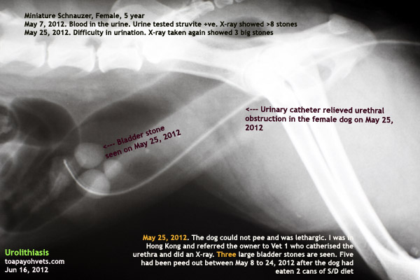
An X-ray of a Miniature Schnauzer that has urethral catherisation. Digital palpation and insertion of a soft catheter was done by Vet 1 as I had referred the owner to her. I was in Hong Kong at that time and the dog had difficulty peeing after passing out some stones. Did the dog owner agree to Vet 1 removing the urinary stones? No.

"Why?" I asked Vet 1 later when I returned to Singapore. She was willing to spay and remove the urinary stones at one surgery whereas I did not want to do two-in-one as I don't increase my anaesthetic and surgical risk by doing a prolonged surgical procedure.


She did not know the reason. "Is it the cost?" I asked her.


"No," she said. "The owners were agreeable to do the two surgeries when they consulted me."


First X-ray shows numerous stones. Advice to remove the bladder stones not taken up as the owners wanted a spay to be included at one surgery, i.e. 2-in-one operation.
Subsequently, second X-ray with urethral catherisation. I checked out this X-ray 24 hours before the spay
PHOTOS ARE IN TOA PAYOH VETS' WEBSITE


Vet 1's boss tries to send image to me via his Blackberry phone.


Owner shows me the 5 stones peed out

Spayed by me

"How much did you quote?"


"The usual rates," she would not give me the actual figure. A fuzzy reply.


"Did you do it immediately?" I asked.


"No, it was a Saturday and the cost would be much higher. I hospitalised the cat. They took back the cat on Sunday."


"Did you follow up?" I asked.


"Yes, but there was nobody answering the phone." This is a good vet as many vets don't do follow up.


"I am sure it is the cost," I said.


"No," she replied. "They had agreed to the surgery."


Other than cost, what could it be? Preference for a veteran?

Later the husband came to me to get the dog spayed. The husband would not give me the reason as to why they did not accept the combined surgery as that was what they wanted. A fuzzy reply saying that the dog could pee after the catherisation. Besides the dog could pass out the 5 stones. So, they did not want to operate. The husband asked: "Vet 1 says the bladder will rupture if I don't remove the bladder stones as they will increase in size. Is it correct?"

"The bladder will rupture if the urine can't flow out for a long time," I said. "The stones will irritate the bladder causing bleeding. As to whether the stones will rupture the bladder by themselves, I don't think they will as they are not sharp." It is a difference of opinion. Some vets will not agree with me.

The dog still has the 3 stones and apparently had no blood in the urine. I did not advise further as this would be like high-pressure selling. The husband had associated feeding 2 cans of S/D diet with the passing out of 5 stones (see image) and got another 6 cans. I had advised 1-3 months feeding of S/D diet for struvite stone dissolution but that means more than 6 cans!

Knowing the reasons of the client is important to improve the standard of care and service. If the vet does not bother to ask, the answers will never be known and the standard of care cannot be improved.

UPDATE: As at Jun 25, 2012, no complaints of blood in the urine. I have not phoned yet as it seems to be "pestering" if the owner has decided not to operate. The spay op was OK otherwise I would have heard from the owner. Since the stitches were dissolvable, I did not see the dog post-op to remove the stitches.
The first report of this case is at:

http://www.sinpets.com/dogs/20120246bladder-stone-spay-packaged-deal-singapore_ToaPayohVets.htm

Cat with rectal prolapse

I presume you are a veterinary foundation. You can use the image in its original form with "toapayohvets.com" in it. Best wishes.


On Sat, Jun 23, 2012 at 9:45 AM, Hannah Lieberman wrote:


Wow, thank you for getting back to me so quickly. That's exactly the one I want. It's very clear.


If we wind up using it, I will absolutely give you credit as requested. I will also send you the link to the finished product.


Oh jeez - I just re-read what I had written. I'm sorry. What I meant to say was that we don't treat rectal prolapses. Because we get so many requests, and because the general public doesn't typically understand what is happening with their pets, I am making lists of what we do and what we don't do, and trying to provide photographic examples for everything on both lists. The rectal prolapse is something that falls under surgeries we don't do. (Sadly.) I hope that doesn't change the status of your permission.


I'm attaching the requested image. Thank you again.


Sincerely,

Hannah Lieberman

Mobile Clinic Director

The Sam Simon Foundation


Use your tools of the trade - opthalmoscope

Yesterday, Friday, Jun 22, 12, I allocated cases to Dr V and Dr D so that waiting times don't drag on as most Singaporean pet owners hate to wait at the Surgery. This is a common trait and many vets and doctors at Clinics don't realise that trait. So, I install this system.

The new client went to Dr V.

"I just want a second opinion," the lady in her 40s said to me. "My vet (many years of experience and consulted by this owner for around past 20 years) said my dog's blindness is due to brain problem and not cataracts. He said there is no cure. MRI may detect where the problem occurs in the brain."

I went into the consultation room to particpate.  After fluorescein stain showed negative corneal ulcers. The dog banged his head when he walked and so there was blindness in both eyes.

WHAT TO DO?
"Did your vet do an eye check with this opthalmoscope?" I asked the lady who came with her brother-in-law whom I mistook for her husband.

"No," she said. I guess her regular vet finds that it is not necessary to use the opthalmoscope or there are other reasons as each vet approaches a consultation differently from others.


NOTES: This may be a case of SARD
Sudden acquired retinal degeneration (SARD)
Bilateral. Middle aged. Older dogs females predisposed. All breeds affected.
Complete blindness
Cause unknown
No treatment
Bilateral absence of the menace and dazzle response
Resting mydriasis
Retina degeneration
Hyper-reflectivity of tapetum
Vascular attenuation
Differential diagnosis:  Central (neurologic) blindness or peptic neuritis
Test: Electroretinogram


This case is a female Maltese, 8 years old, completely blind. Will wait for blood test report. From the opthalmoscopy, I saw in both eyes, the lower half of the fundus being very shiny reddish brown (hyper-reflectivity) like semi-circles. The upper half is greyish white with bigger blood vessels (Vascular attenuation) seen. The optic disc appears swollen (oedematous) - optic neuritis?

Follow up: Rabbit with large cheek abscess


E-MAIL TO DR SING DATED Jun 19, 2012
Dear Dr Sing,

here are the photos of Huihui and Ban Ban's most recent photos. No 18 is to let you see how they are housed currently, normally no partition only cos Huihui needs to be separated from Banban currently.

Thanks a lot for helping explain many things to us also and all the advice and recommendations.

Just to inform you that Huihui has started eating the pellets slowly and had drank at least 10mls of apple juice.


E-MAIL TO DR SING DATED Jun 24, 2012

I want to ask when should I stop pumping in the chlorohexidine wash into his face as the hole actually closes up every morning and evening and I have to poke through the sealed hole each time I wash..... The swelling has gone down considerably but the area around the hole is quite hard so I am not sure if there's anything wrong. I am not sure if there is still any pus inside although I thought I see some whitish stuff inside. His appetite has gone back to normal and his speed of eating has returned to normal. He also started to lick me after each treatment, which I considered as a good sign as he hasn't did that since the swelling started. He has also regained his energy, running and jumping up and down his toilet, standing up frequently to beg for treats/pats. So can you please advise me if I should continued poking through the hole to wash or just leave the hole to closed up?

Sorry for the long letter but I thought I should let you know how he is doing


E-MAIL FROM DR SING DATED Jun 24, 2012

I am glad to see one of your two rabbits after over 5 years. I am still checking my website the original picture taken when I neuter and spay your two rabbits.

You have written an excellent post-op nursing report. Good detailed report from the owner is very rare and is useful as a follow-up on the operation (lancing of the abscess done by Dr Vanessa before the X-ray as advised by me).

You need to flush out as much pus from deep inside the abscess and therefore continue irrigation till you don't see any "white" pus inside the cavity.

1. From the X-rays, the abscess from the elongated impacted molar is very deep and molar tooth extraction should be the solution to prevent recurrence of the abscess. However, this procedure is not so easy in a rabbit compared to the dog and cat.

2. The rabbit needs to be checked for molar spurs every 4 weekly. Trimming of the spurs to the gum line may be necessary in malocclusion.
3. Make an appointment next Sunday morning July 1, 2012 to check on the molars and the abscess. I am in the midst of writing a case report of your rabbit X-ray. It takes lots of time to do it. Best wishes.

Saturday, June 23, 2012

1047. Permission to use image of cat with rectal prolapse

On Sat, Jun 23, 2012 at 5:52 AM, Hannah Lieberman wrote:


Hi there,

I run a non-profit foundation mobile veterinary clinic that we take to low-income neighborhoods in Los Angeles, CA three days a week to perform veterinary surgeries on pets belonging to low-income residents. All services are completely free.


I am currently building a new website, and including photos of ailments that we're able to treat in our clinic. Since we treat rectal prolapses, I'd like to include a picture of one.

I really like the picture of the cat with the rectal prolapse you have your website, and would like to include it on ours. May we get permission to do that, please?

E-MAIL REPLY FROM DR SING DATED JUN 23, 2012
I am Dr Sing from Toa Payoh Vets. Which picture hyperlink is that? Please let me know.


You can use the picture of the cat with rectal prolapse if you use it without deleting "toapayohvets.com" acknowledgement. Good luck.


Friday, June 22, 2012

1046. Closed Pyometra - blood tests and x-rays

Golden Retriever, Female, 10 years

Not eating for 4 days. Lethargy. No fever.

History: Polydipsia & Polyuria

1.  Blood test:
Total WCC = 38 (6-17). N86%, L4%, M9%, E1%, B0%

2. X-rays: Uterine horns filled with pus
3. Ultrasound: Pus seen in both uterine horns. Cervix is closed.

Diagnosis: Closed pyometra

Surgery: 1 hour. Spayed.

Outcome: Good. Dog is back to normal.

High total white cell count and history indicated closed pyometra. X-rays and ultrasound are useful to confirm pus in the uterus.

1045. Example of a veterinary report


From Jun 23, 2012, all veterinary surgeries will be recorded in the following format:

http://www.bekindtopets.com/animals/20120613veterinary-report-format-adenocarcinomasingapore-ToaPayohVets.htm


\

From Jun 23, 2012, all vets at Toa Payoh Vets will record the veterinary surgeries done in the following format and e-mail to Dr Sing. This e-mail will be stored in the "cloud". An example is given below:


Veterinary surgery report - excision of bladder adenocarcinoma on May 14, 2012 & follow up 38 days later on Jun 21, 2012


Date of Veterinary Surgery Report: Jun 22, 2012

Name of Veterinary Surgeon: Dr Sing Kong Yuen & Dr Daniel Sing


Case seen on May 14, 2012 by Dr Daniel Sing

1. History & Complaint: TP 43355, (Name of Dog), Beagle M, 13 years, attended by Dr Daniel Sing


2. Presenting Sign & Symptoms: Incontinent with bloody urine dripping all over the floor and passing blood clots for > 4 weeks. Haematuria + Anaemia


3. General Examination: Abdominal pain


4. Tests done & significant findings of tests:

4.1 Blood tests

May 10, 12. RBC low 5.1 (5.5-8.5), Total WCC 12.4 (6-17), N 75%, L12%, M7%, E5%,B1%

Jun 21, 12. RBC 5.6 (5.5-8.5), Total WCC 12.1 (6-17), N 77%, L16%, M7%, E0%,B1%


4.2 Urine tests

May 10, 12. pH 5, SG 1.005, Protein 3+, Ketones 1+, Blood 4+, WBC 90, RBC>2250, Bacteria 3+


Jun 21, 12 pH 7, SG 1.008, Protein 1+, Ketones -ve, Blood 3+, WBC 48, RBC 351, Bacteria 3+


4.3 Histopathology: Moderately differentiated adenocarcinoma


5. X-rays: Radio-dense "stones" seen in bladder area.


6. Anaesthesia: May 15, 12. Body weight: 16 kg. At 50%, Dom 0.32, Ket 0.4 ml IV. Isoflurane gas maintenance. Surgery time: 2 hours. Polysorb absorbable sutures 2/0 x 3 packets, 3/0 x 1 packet


7. Surgery: May 15, 12. Excision of hard gritty tumour, 1.8cm x 1.5cm x 0.5 cm, near the neck of the tumour. Sent for histopathology. Operating surgeon: Dr Sing K Y. Assisted by: Dr Daniel Sing


Follow-up dates: May 22, 12 7.35 pm, Dr Sing K Y phoned owner. No response.

Jun 22, 12, Dr Daniel Sing informed owner of the 2nd blood and urine test results


Signature of Veterinary Surgeon: Dr Sing K Y

Date of this report: Jun 22, 2012


Attached and owner has acknowledged receipt:

Blood tests given to owner: No

Urine tests given to owner: No

X-rays given to owner: No

Ultrasound report given to owner: Not Done.

Histopathology report given to owner: No

Sterilisation Certificate No. .... given to owner: NA

Others given to owner: NA


Anaesthetic & Surgical Record No. ........(Form is not printed yet).

Ear Scope: NA

Skin scraping: NA

Opthalmoscopy: NA

Urinary stone analysis report: NA

Others: NA


Comments by Dr Sing K Y

Another two attempts to phone the owners were not successful. On Jun 21, 12, the owners came with the complaint of painful left hip and re-stocking of anti-seizure medication which must be given daily to prevent seizures. Jun 21, 12 was 38 days after the surgery.


Outcome of surgery: Good. On Jun 21, 12, the couple is very happy that the dog is active and no longer drips dark brown blood and passes blood clots everywhere. They said the urine is clear and has no blood. The dog is able to control his bladder during the daytime but not at night. The dog weighed 15 kg compared to 16 kg during first visit but is putting on weight. Bladder palpation by Dr Sing elicited a painful whine from the Beagle. The bladder was around the size of the tennis ball. Catherisation by Dr Daniel Sing did not produce urine. The wife came later in the afternoon with a bottle of urine collected directly from the dog.


Results: This 2nd urine test indicated UTI and demonstrated the presence of blood. The lab reports will be given to the owner at the next visit.


2nd urine test: Jun 21, 12 pH 7, SG 1.008, Protein 1+, Ketones -ve, Blood 3+, WBC 48, RBC 351, Bacteria 3+


baldder adenocarcinoma seen during surgery. beagle, male, 13 yearsCase report at Toa Payoh Vets:

tpvets_logo.jpg (2726 bytes)5301.

Trust & Audit in a Case of an old Beagle with fits and bladder cancer


http://www.sinpets.com/dogs/20120440veterinary-audit-cystotomy-carcinoma-bladder-toapayohvets-singapore_ToaPayohVets.htm