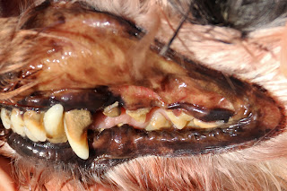
A 12-year-old male neutered Miniature Schnauzer had not been eating for a week. He was certified fit to travel from the USA to Singapore 3 months ago. He had been "coughing" phlegm for some days. He is not on medication including steroids.
He was fed a BARF (Biologically Appropriate Raw Food Diet) for around 2 months and then home-cooked chicken diet for the past month. Dog treats were given.
Physical examination
Thin, no fever. No icterus. Heart sounds muffled. Slight anterior abdominal pain. No abdominal masses were palpated.
Radiography
The heart and liver are not enlarged. The liver size and shape are normal. No large abdominal masses.
Blood test.
1. High total cholesterol. Triglyceride level is normal.
2. Low fasting glucose
3. High ALT and AST (markers of hepatocellular damage). Alanine aminotransferase (ALT) and aspatate aminotransferase (AST) are liver enzymes.
Total cholesterol 22.4 mmol/L (3.5 - 7.2)
Triglyceride 1.8 mmol/L (0.3 - 3.3)
ALT 3722 U/L (10 - 109)
AST 587 U/L (13 - 15)
4. No leucocytosis or leucopaenia. Unlikely to have leptospirosis or viral infections but this cannot be ruled out.
5. Kidney function is normal
Ultrasonography (focal liver lesions, diffuse liver disease or biliary disease like cholestasis), evaluation of urine or serum bile acids and liver biopsy (fine needle aspiration, laparoscopy, exploratory laparotomy) were not done owing to financial constraints.
Conclusion
An increase in total cholesterol, ALT and AST liver enzymes indicate liver disease in the dog.
Persistent ALT increases should be investigated when they are greater than twice normal. The most important diagnosis to make is chronic hepatitis. Early diagnosis and prompt therapy improves patient survival
Some cases of liver failure can be reversed. The causes can be hepatic or non-hepatic .
The laboratory tests must be correlated with the history, physical examination to arrive at a diagnosis.
In this case, the dog is not overweight. The high total cholesterol might be due to the diet including dog treats.
Miniature Schnauzers are a breed predisposed to liver disease during old age. A common condition in the older dog is idiopathic vacuolar hepatopathy resulting in elevated ALP.
In conclusion, 4 categories of causes of elevated liver enzymes in the dog are:
1. Primary hepatobiliary diseases.
2. Secondary to extra-hepatic diseases
3. Benign condition (hepatic nodular hyperplasia)
4. Progressive condition (chronic hepatitis, neoplasia).
Advices to the old dog owner
Repeat blood test in 4 weeks. If liver enzymes are elevated, further tests are needed (ultrasonography, bile acids, wedge liver biopsy) to check for chronic hepatitis, hepatic neoplasia, benign hepatic nodular hyperplasia and diffuse vacuolar hepatopathy
.
Low fat high fibre quality maintenance diet. Hand-feeding is important.
Liver support therapy eg. Zentonil
VIDEO
24 HOURS after treatment
Day 3 of treatment. Mild jaundice is seen in the eye sclera which is stained very light yellow.
Day 4 of inpatient
Can stand, walk, poop. On I/V drip. Not eating the K/D and A/D today. Hand fed. Urine testing.
Eye sclera looks more yellowish.
--------------------------------------------------------------------------------------------
Hyperlipidemia is a condition in which the amount of fats (also called lipids) in the blood are elevated. The most important lipids are cholesterol and triglyceride. Hyperlipidemia is a common and under-diagnosed dog health problem that can negatively impact health and longevity.
Several metabolic diseases demonstrate hyperlipidemia, including diabetes, hypothyroidism and Cushing’s syndrome. Some dog breeds are genetically predisposed to hyperlipidemia.
Hyperlipidemia does not normally lead to heart disease, but can decrease lifespan and cause obesity, neurologic and metabolic issues.
Genetic predisposition – Miniature schnauzers and Beagles tend to be genetically predisposed to hyperlipidemia.
Symptoms of hyperlipidemia can include: Decreased appetite Vomiting Diarrhea Abdominal pain Bloated abdomen Cloudy eyes Fatty deposits under the skin Hair loss Itching Seizures
Diagnosis of High Cholesterol in Dogs You may want to eliminate all table scraps and gradually switch your pet over to a low-fat, high-fiber dog food as diets high in fat are a common cause of hyperlipidemia. However, results from diet changes can take 6-8 weeks.
If you are seeing symptoms associated with hyperlipidemia in your pet, you will need to visit the veterinarian to determine the underlying cause.
A full history of your pet and a thorough physical exam will determine what diagnostic tests may be necessary.
-----------------------------------------------
The common bile duct is a small, tube-like structure formed where the common hepatic duct and the cystic duct join. Its physiological role is to carry bile from the gallbladder and empty it into the upper part of the small intestine (the duodenum). The common bile duct is part of the biliary system.
Bile is a greenish-brown fluid that helps digest fats from our food intake. It is produced by the liver and stored and concentrated in the gallbladder until it is needed to help digest foods. When food enters the small intestine, bile travels through the common bile duct to reach the duodenum.
Gallstones are hard deposits that form inside the gallbladder when there is too much bilirubin or cholesterol in the bile. Although a person may have gallstones for many years without feeling any symptoms, gallstones can sometimes pass through the common bile duct, causing inflammation and severe pain. If a gallstone blocks the common bile duct, it can cause choledocholithiasis. Symptoms of choledocholithiasis include pain in the right side of the abdomen (biliary colic), jaundice, and fever. Choledocholithiasis can be life-threatening if not diagnosed and treated immediately.
INTERPRETING LAB TEST RESULTS ON RABBITS
Understanding those emailed or printed results we get after our rabbit’s lab test can be difficult. I hope that the following explanations of two commonly‐ordered tests will help readers interpret their rabbit’s test results.
Printed results of the tests may be presented in graphical or numerical form. They usually include the test name and/or abbreviation, results, and the low and high ends of the normal range for that test.
Rarely, errors in taking, handling or analyzing the sample will cause erroneous results, particularly if the sample is not stored correctly or is not analyzed soon enough after taking it. If you have concerns about the validity of a test result discuss it with your vet. In some cases a new test may be warranted.
Haematology.
In a CBC, or complete blood cell count, the amounts of the different kinds of blood cells present are tested.
Red blood cell (RBC or erythrocyte) count: Male rabbits and older rabbits tend to have higher counts than female and younger rabbits. Dehydration and stress from cold temperatures can cause high RBC counts. High counts of nucleated RBCs can be a sign of a bacterial infection; a very high count of nucleated RBCs can be a sign of a bad flea infestation or internal bleeding.
Slightly elevated counts of nucleated RBCs are not an abnormal finding in rabbits. The HCT (Hct, haematocrit) is a test in which the percent of red blood cells is calculated. A low value may be a sign of anemia. Hb, or hemoglobin concentration, can be used to help diagnose anaemia (low Hb) and its origin. Female rabbits tend to have much lower HB and Hct than males. Rabbits that get a lot of exercise may have elevated RBC, HB, and Hct values.
Platelets: High counts may be associated with iron‐deficiency anaemia and chronic bleeding. Cold stress can also cause elevated platelet values, as may drugs such as glucocorticoids and epinephrine.
White blood cells (WBC, leukocytes): White blood cell counts vary depending upon the age, sex, breed and season of the year.
Serum/blood chemistry. The focus on these tests is on parameters of blood other than cell counts.
Serum glucose: Although high glucose can be a sign of kidney disease, it is often caused by stress, including the stress of the trip to the vet and having blood drawn. High glucose values can also occur in rabbits with acute intestinal obstruction, hepatic lipidosis, hyperthermia, and shock.
BUN (blood urea nitrogen): A key test used to assess kidney function. Urea levels depend upon a wide variety of factors, including the time of day, the amount of protein in the diet and how hydrated the rabbit is. Slightly high values are not uncommon in healthy rabbits.
Creatinine: High values are often a sign of severe kidney or muscle damage. This test is less influenced by external factors than the BUN. However, levels may be high in rabbits that have gone a few hours without drinking water. A disadvantage of this test is that it does not show high levels until there has been substantial loss of kidney function (excepting temporarily high levels caused by dehydration, which do not involve such loss of function).
Cholesterol and triglycerides: To obtain accurate values for cholesterol the animal must be fasting. Since it is unsafe to fast rabbits for more than a couple of hours, not to mention that it is essentially impossible to fast most rabbits because of consumption of cecotrophs, results from this test should be considered only in conjunction with other tests.
Calcium and phosphorus: Calcium levels are primarily influenced by the calcium content of the diet. High blood calcium levels in conjunction with clear urine (showing it is not being excreted as it should) are a sign of kidney failure.
Serum protein: Total protein levels may be high in rabbits that are dehydrated, whether from gastrointestinal hypomotility (stasis) or other reasons. Low levels can be caused by malnutrition or liver disease. Low levels of albumin (a specific protein) can be a sign of a heavy infestation of parasites, and high levels are a sign of advanced liver disease.
Bilirubin: High serum bilirubin levels in young rabbits are often caused by hepatic coccidiosis; in older rabbits they are more likely to be caused by an obstruction of the bile duct by neoplasia (cancer) or an abscess.
AP (ALP, alkaline phosphatase): The normal range for this test is wide and varies with age (young rabbits have higher levels) and breed. A high value may be a sign of diseases affecting liver function such as hepatic coccidiosis, liver abscesses, and neoplasia.
ALT (alanine aminotransferase), also called GPT and SGPT: Another test that helps the vet assess whether there has been any liver damage. Mildly high levels may be found in rabbits that appear healthy and it is thought they may be caused by low concentrations of toxins such as aflatoxins in food or compounds in wood‐based litters.
AST (aspartate aminotransferase), formerly called SGOT: High values in conjunction with high ALT, AP, or protein may be a sign of liver damage. High levels may also be caused by the rabbit struggling during collection of the sample.
Urinalysis
The urinalysis is another lab test that is ordered fairly frequently. More so than in blood work the normal value is often “negative,” or the absence of the tested‐for compound.
Protein: The normal finding is negative to trace amounts. High amounts can be a result of kidney damage/disease. High levels can also be caused by dehydration, strenuous exercise or stress, and for this reason protein is best looked at in conjunction with the specific gravity and the ratio of protein to creatinine. Dilute urine with a high protein value is more likely to be a sign of kidney damage/disease than concentrated urine with a similar protein value. Low protein levels can be a sign of malnutrition.
SG (specific gravity): An SG level at the lower end of the normal range when combined with high creatinine and BUN is a sign of poor kidney function.
pH: Normal rabbit urine has a high pH (7.5‐9). Lower pH can be caused by high‐protein diets, severe
anorexia, and fever.
Glucose: The normal finding for glucose is negative, although trace amounts may be found in healthy rabbits. Higher amounts can be caused by stress, including pain or any experience which is frightening to the rabbit.
Ketones: The normal finding is negative. The presence of ketones indicates starvation or severe anorexia. Ketones may be seen in rabbits with severe dental disease that prevents them from eating or in rabbits on hay‐only diets that have severely impacted cecums. Rarely, ketones in the urine may be caused by diabetes mellitus.
Bilirubin: High levels of bilirubin in rabbit urine are unusual but elevated levels may be caused by poisoning from aflatoxins in contaminated food, hepatic coccidiosis, or neoplasia.
Haematuria: Normally there is no blood present in rabbit urine. A positive result may be caused by inflammation and/or a urinary tract infection, crystals, or, less often, neoplasia.
Sediment: Calcium carbonate sediment is a normal finding in rabbit urine. Other crystals can be caused by drugs the rabbit is taking, and struvite crystals can be a sign of bacterial infection.
Urobilinogen: The finding for a healthy rabbit is negative. High levels may be caused by liver damage or drugs such as sulfonamides; low levels can be caused by long exposure to light.
Nitrate and nitrite: Urine normally contains nitrates, but some bacteria convert nitrate to nitrite. The normal result for nitrite is negative; a positive result is caused by bacteria in the urine. However, not all bacteria are able to convert nitrates to nitrite, so a negative result does not mean a UTI is not present.


No comments:
Post a Comment
Note: Only a member of this blog may post a comment.