Wednesday, September 2, 2020
3410. Leishmaniosis - zoonosis - notifiable disease
Leishmaniosis
Leishmaniosis is a vector-borne zoonotic disease caused by the Leishmania spp. parasite.
Transmission to animals including dogs and humans is primarily through the bite of infected sandflies.
Direct dog-to-human transmission has never been reported worldwide. The disease is most prevalent
in tropical and sub-tropical regions, mainly in Africa, parts of Asia, the Middle East, Latin America
and the Mediterranean region.
2 Clinical signs in dogs are variable and can mimic other illness. Some of the signs and
symptoms may include skin lesions, weight loss and organ failure. Infected dogs may not always
show signs. The incubation period in dogs can range from months to years. Chemotherapeutics are
available for the treatment of this disease.
Treatment for leishmaniosis can improve the condition of
the dog, but it may not eliminate the parasite. In such cases, an infected dog may remain a carrier of
Leishmania species.
Advice on protecting animals from leishmaniosis
3 To reduce the chance of being bitten by sandflies, owners and carers can apply topical insect
repellent on their dogs or use collars that are impregnated with insect repellents (e.g. deltamethrin)
against sandflies. It is also advisable to avoid visiting areas where there are sandflies during dawn
and dusk and practice good personal hygiene such as washing of hands when caring for their dogs.
Advice on the handling of Leishmania cases in animals
4 Due to the fact that infected and previously infected animals remain a potential risk to public
health and the health of animals, we would like to advise on the measures to take to minimise the
risk of transmission of Leishmania for any suspect or infected animals:
a. Housed in a vector-protected area to avoid contact with other dogs and sandflies, where
possible;
b. Measures should be taken to avoid being bitten by flies; e.g. avoid going out at dawn and
dusk or apply an insect repellent collar or spray;
c. NParks/AVS should be informed if other dogs are to be kept on the same premises;
d. Bodily waste should be properly disposed of;
e. People coming into contact with these animals should practise good personal hygiene, such
as washing their hands after interaction;
f. Immuno-compromised individuals should avoid contact with these animals;
g. Undergo maintenance treatment for leishmaniosis;
h. NParks/AVS must be informed whenever there is a change in the health status related to
leishmaniosis;
i. Any new attending veterinarians should be informed of the animal’s history of leishmaniosis;
j. These animals must not be used as a blood donor and breeding of the animal should be
avoided.
Sunday, August 30, 2020
3409. STOCK PHOTOS FOR SALE by Dr Sing Kong Yuen. www.shutterstock.com/g/toapayohvets
UPDATE AS AT 1 NOV 2022:
All
stock photos of Singapore buildings, people, flora, fauna for sale, scenaries and other countries are at:
www.shutterstock.com/g/toapayohvets
Contact: Dr
David Sing
+65
9668-6468
judy@toapayohvets.com
99pups@gmail.com
www.toapayohvets.com
This
webpage
https://2010vets.blogspot.com/2020/08/3409-singapore-wildlife-seen-on-30-aug.html
A few examples of stock photos are shown below:


3408. Histology report for tumours
After tumour removal, we send the tumour for histology. The cost is from $350 to $400.
With histology, we know whether the tumour is cancerous or not.
For example, this 7-year-old male Golden Retriever has a gigantic elbow tumour. At first, the owner thought it was the common elbow hygroma as displayed during internet research. Hygromas are not cancerous. This case is a poorly differentiated sarcoma. There is recurrence after surgical removal.
The details are at:
https://2010vets.blogspot.com/2020/07/4131-7-year-old-golden-retriever-has.html


Saturday, August 29, 2020
3407. Governor's House. Pyin Oo Lwin in 2020
9 Sep 2020.
MAKING A VIDEO ABOUT THE GOVERNOR'S HOUSE
I am Dr Sing Kong Yuen, veterinary surgeon from Singapore, Toa Payoh Vets, www.toapayohvets.com.
I enjoyed my stay in the Governor's House-the biggest room sometime in 2006-2008. I was probably the only foreigner. I stayed in the Governor's room. The ambience was excellent and the service was excellent. The managing director was a nice man in his late 70s. He gave me some history of this place. He was a retiree helping out the founder of this resort. It is 2020 now and a decade has passed. Now you have beautiful surroundings.
FOR IMPROVEMENT TO YOUR SALES
I notice that your presentation is more of the same "luxury" resort facilities. While they are important, they are also present in other luxury resorts. Your unique selling proposition is the HISTORY of the British colonial era - the various governors. You have a plaque inside your hotel with the names of the British Governors. Is there really a museum?
I am making a video about the Governor's House. Will it be possible for you to allow me to use the images in this webpage and provide me more information of your museum and the horse and carriage tours? This video will help to inform guests who may have high expectations of this resort understand better. The high expectations from a Singaporean guest and others are recorded in the google reviews.
Thank you very much.
Dr Sing Kong Yuen.
My tel is +65 9668-6468, 99pups@gmail.com, Singapore Toa Payoh Vets.
------------------
The Aureum Resort is comprised of 34 spacious, en suite bungalows scattered through the beautifully manicured gardens surrounding the Governor’s House.
The style is as you expect; traditional with classic furnishings, but with all the modern in-room comforts and amenities. There is also a presidential suite available.
Facilities & services
- 34 bungalows
- 5 governor's suites
- 1 presidential suite
- Restaurant
- Indoor & outdoor swimming pools
-------------------------
-------------------------
Florian Schulz9 months ago on  Google
Google5/5
====================
IMAGES AT:
======================


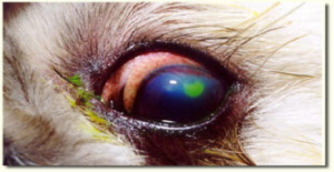
3406. A Labrador Retriever puppy in Myanmar has an eye growth - ocular dermoid.
HOOK
 |
| A normal Labrador Retriever puppy is born with clear eyes. There are no eye growths irritating him. |
But this 10-week-old Labrador Retriever is unfortunate. His right eye has a fatty growth with hairs since he was born.
 |
He tries to relieve his itch by rubbing his eye with his paw or on his bed mattress.
The upper eyelid area is bald. This is due to continual rubbing.

The Owner is very worried. He consulted Dr May Than Oo, a veterinarian in Pyin Lwin Oo. This Labrador Retriever lives in Pyin Oo Lwin.
PYIN OO LWIN
is a scenic hill town with a cool climate around November to February. It is a tourist destination. It was a colonial town for the British Governor to stay during the hot season in Yangon many years ago. The Governor's House was destroyed during the 2nd World War. It is now rebuilt and is a luxury hotel.
What is this fatty growth?
It is an Ocular Dermoid.
As it is located in the conjunctiva, it is classified as a lipodermoid.

A dermoid is a growth containing normal skin parts. These parts include the epidermis, dermis, fat, sebaceous glands, hair follicles, and frequently hairs.
In dogs, Ocular Dermoids most frequently locate at the temporal limbus but can involve the conjunctiva, eyelid, nictatating membranes, limbus or cornea. In this case, the ocular dermoid is at the temporal conjunctiva and does not involve the limbus or cornea.
How to get rid of it?

An operation to cut it out is needed. It is called surgical excision.

After excision, the upper and lower eyelids are sewn up to facilitate healing and an Elizabeth collar is worn for around 14 days.
 |
This is a case from the Veterinary Files From Myanmar, sponsored by Toa Payoh Vets. OCULAR DERMOID IN THE DOG
How to get rid of it?
This video discusses the surgical treatment of an ocular dermoid
This video contains surgical procedures.
VIEWER DISCRETION IS ADVISED.

----------------------------------------------------

-----------------------------------------------------------------
A third-eyelid flap was also performed to facilitate healing. After surgery, eye drops as atropine sulfate, gentamicin sulfate, diclofenac sodium and systemic antibiotics were prescribed for 2 weeks.
After 2 weeks, the third-eyelid flap was removed, and the region of conjunctival flap was epithelized and remained the scar
Signs - epiphora, pigmentary keratitis, ulcerative keratitis
------------------------------------------------
Visuals are presented:
1. An ocular dermoid causes epiphora and eye irritation.

2. The dermoid is pulled up using two forceps.
 |
| Two forceps clamp and pull up the dermoid.
3. Not shown in this video: The base of the dermoid is clamped with forceps.
|


5. The incised conjuctiva is not stitched. Its wound will heal. There is no need for a
conjunctival flap.

6. The third-eyelid flap procedure is used to cover the eyeball. This will facilitate healing of
the conjunctiva as it is not irritated by external dust or air currents. The upper and lower eyelids
are brought together using tubings for 14 days.
are brought together using tubings for 14 days.


7. Antibiotic eye drops or ointment are applied daily

Oral Painkillers are given

post op video footage
CREDITS
Special thanks to: Dr May Than Oo for the images of the surgical excision.


FOR MORE INFORMATION

-----------------------------------------------------------------
RESEARCH NOTES
DIFFERENT TYPES OF OCULAR DERMOIDS
In dogs, Ocular Dermoid most frequently locate at the temporal limbus but can involve the conjunctiva, eyelid, nictatating membranes, limbus or cornea. A Shih Tzu has a dermoid - electro-excision case study
In dogs, Ocular Dermoid most frequently locate at the temporal limbus but can involve the conjunctiva, eyelid, nictatating membranes, limbus or cornea. A Shih Tzu has a dermoid - electro-excision case study
LOCATIONS OF THE OCULAR DERMOID.
1. A cat keeps rubbing his eyes. Why?He has a limbal dermoid.
Show footage 0:00 to 2:04. No need to edit audio. Just insert a slide saying:
"In this case, the dermoid is present on the limbus (sclera and cornea junction).
WHAT IS A DERMOID?
A dermoid is normal skin as it has epidermis, dermis, fat, sebaceous glands, hair follicles and hair. The tissues are usually irritating the eye Thus, the patient suffers from chronic epiphora (excessive eye tears dropping and keratitis (inflammation of the cornea).
A dermoid is an overgrowth of normal, non-cancerous tissue in an abnormal location. Dermoids occur all over the body. The ones in and around the eye are usually comprised of skin, hair, and/or fat.
WHERE ARE DERMOIDS FOUND AROUND THE EYES?
There are two main dermoid types that occur on or around the eyes.
First, an orbital dermoid is typically found in association with the bones of the eye socket.
Second, an epibulbar dermoid is found on the surface of the eye. There are two typical locations for an epibulbar dermoid. One of the locations is at the junction of the cornea, the clear part at the front of the eye, and the sclera, the white part of the eye. This is a limbal dermoid. The second location of an epibulbar dermoid is on the surface of the eye where the lids meet in the temporal corner (towards the ear) which is often called a dermolipoma or lipodermoid.

The lesion invaded the stromal layer of the cornea, and extended to limbus and conjunctiva. It was surgically resected.
*A pedicle conjunctival flap was placed to support the reepithelization and aid vascularization of the defect.
*A pedicle conjunctival flap was placed to support the reepithelization and aid vascularization of the defect.
*After excision, the site where the dermoid lay can be covered by a piece of transplanted cornea.
A third-eyelid flap is also performed to facilitate healing.
After surgery,
An Elizabeth collar to be worn for 2 week.
Eye drops with antibiotics and systemic antibiotics are prescribed for 2 weeks.
*needs operating microscope
*needs operating microscope
Ocular dermoid in a cat.
Treatment: Surgical excision using Surgical Blade No. 11.
The lesion invaded the stromal
layer of the cornea and the sclera at the limbus. The cat was not operated as the owner did not give consent since this was a "stray" cat.
----------------------------------------------------------------------------------
2. A Shih Tzu has a corneal dermoid.
No need to edit. Include the whole footage.
A shih tzu has dermoid in the left eye
----------------------------------------------
3. A labrador retriever has a limbal dermoid.
Give the video link and show some footages of the surgical procedures.
---------------------------------------------------------------------------------------------Give the video link and show some footages of the surgical procedures.
Removal of dermoid by superficial keratectomy is
essential to relieve the related clinical signs (chronic keratitis and epiphora, eye irritation). If the dermoid
has not been totally removed, some degree of recurrence can
be expected [1]. Thus, the dermoid have to excise
completely, if possible, without scarring of the cornea. Once
corneal epithelization is complete, as evidenced by the lack
of fluorescein retention, topical antibiotic-corticosteroid
preparations can be administered to reduce postoperative
corneal scarring and improve the eventual transparency of
the cornea.
After fixation of the globe, abnormal tissue at the conjunctiva
and cornea was removed using the blade (No. 11) and
microsurgical instruments. The lesion invaded by stromal
layer of the cornea, and extended to limbus and conjunctiva
was surgically resected. And then, a pedicle conjunctival
flap was placed to support the reepithelization and aid
vascularization of the defect. Additionally, third-eyelid flap
was also performed to facilitate healing.
-------------------------------------------------------------------------------------------------------
RESEARCH NOTES
VETERINARY DERMOID
Surgical correction of corneal dermoid in a dog
Jae-il Lee1
, Myung-jin Kim1
, Il-hwan Kim2
, Yeoung-bum Kim3
, Myung-cheol Kim1,*
1
Laboratory of Veterinary Surgery, College of Veterinary Medicine, Chungnam National University, Daejeon 305-764, Korea
2
R&D Center of Pharmaceuticals, CJ Corporation, Icheon 467-812, Korea
3
Korea Institute of Toxicology, KRICT, Daejeon 305-343, Korea
A five-month-old female Shih-tzu puppy was presented
for repair of congenital choristoma in left eye. The patient
was suffered from chronic epiphora and ocular discharge
during 3 months. On ophthalmic examination, left eye
revealed hyperemia in conjunctiva of the temporal canthus
due to choristoma with hair. At surgery, the choristoma
invaded by stromal layer of the cornea, and extended to
limbus and conjunctiva. Based on the anatomical location
and histopathological features of the removed tissue, the
choristoma was diagnosed as corneal dermoid.
Key words: choristoma, corneal dermoid, epiphora, hyperemia
Corneal dermoid is a congenital choristoma characterized
by the presence of heterotopic cutaneous tissue in an
inappropriate place [8,4]. They may affect the eyelids,
conjunctiva (palpebral and bulbar), nictitating membrane,
and cornea [3]. This condition is known to occur in large
breed dogs such as St. Bernards [1-3, 7], German Shepherds
[1], short-legged dogs [7] such as Basset Hounds, Dachshunds
and Welsh Corgis and cats [5].
Dermoids contain many of
the elements of normal skin such as epidermis, dermis, fat,
sebaceous glands, hair follicles, and frequently there is hair.
The tissues are usually irritating the eye and associated
structures [3]. Thus, the patients have been suffered from
chronic epiphora and keratitis.
Dermoid may be surgically
excised with complete remission of signs and minimal
scarring of the cornea. This paper describes the incidence of
corneal dermoid and detailed histopathological findings in
shih-tzu dogs.
A 5-month-old female Shih-tzu puppy with a weight of
4.4 kg was referred to the Veterinary Medical Teaching
Hospital of Chungnam National University for repair of
congenital choristoma in left eye. The patient had suffered
from chronic epiphora and some ocular discharge during 3
months. On ophthalmic examination, left eye revealed mild hyperemia in conjunctiva at the temporal canthus.
Vital
signs and results of blood examination were within normal
ranges. A light peach color lesion measuring 3-5 mm in
diameter was noted grossly at the limbus in the direction of
5 o’clock, and there was hair growing from the surface (Fig.
1).
The surface of lesion was rough and slightly protruded
compared with the surrounding normal cornea.
The patient was premedicated with atropine sulfate (0.04
mg/kg, SC). Anesthesia was induced with thiopental sodium
(12.5 mg/kg, IV) and maintained with isoflurane. The
patient was administered a balanced electrolyte solution (10
ml/kg/hr, IV), and cefazolin sodium (20 mg/kg, IV) as
prophylactic treatment was administered before surgery.
After fixation of the globe, abnormal tissue at the conjunctiva
and cornea was removed using the sugical blade (No. 11) and
microsurgical instruments.
The lesion invaded the stromal
layer of the cornea, and extended to limbus and conjunctiva
was surgically resected. And then, a pedicle conjunctival
flap was placed to support the reepithelization and aid
vascularization of the defect. Additionally, third-eyelid flap
was also performed to facilitate healing. After surgery, eye
drops as atropine sulfate, gentamicin sulfate, diclofenac
sodium and systemic antibiotics were prescribed for 2
weeks.
For histopathological evaluations, the tissue sample was collected to 10% neutral phosphate-buffed formalin,
processed routinely, and stained with hematoxylin and
eosin.
After 2 weeks, the third-eyelid flap was removed, and the
region of conjunctival flap was epithelized and remained the
scar (Fig. 2). The dermoid hasn’t recurred for 11 months
since the surgical correction, and hyperemia of conjunctiva
and epiphora was disappeared.
Corneal dermoids are ectopic eyelid tissues. They are
nearly always covered with hair. Although, hair may be
removed by manual epilation or electroepilation, it may
regrow. Corneal dermoid has been reported in various
species of animals and in humans, and it is commonly
believed that this disease is generally congenital, although
not hereditary [4]. However, some report in humans, the
appearance of corneal dermoid across three generations of a
single family has been reported by Mattos and his colleagues
[6]. In this case, hereditary pattern was not revealed because
parents were normal ocular structure.
Removal of dermoid by superficial keratectomy is
essential to relieve the related clinical signs. If the dermoid
has not been totally removed, some degree of recurrence can
be expected [1]. Thus, the dermoid have to excise
completely, if possible, without scarring of the cornea.
Once
corneal epithelization is complete, as evidenced by the lack
of fluorescein retention, topical antibiotic-corticosteroid
preparations can be administered to reduce postoperative
corneal scarring and improve the eventual transparency of
the cornea.
Microscopically, it was presented the corneal
dermoid invasive normal corneal epithelium, and the
dermoid contain normal skin such as hair follicles, cornium
and blood vessel (Fig. 3).
The operation of dermoid was delayed in this case due to
owner’s circumstances. Consequently, the lesion was increased
in size and extended more invasively. Fortunately, dermoid
was removed successfully and recurrence did not appear up
to now.
References
1. Gelatt KN. Bilateral corneal dermoids and distichiasis in a
dog. Vet Med Small Anim Clin 1971, 66, 658-659.
2. Gelatt KN. Corneo-conjunctival dermoid cyst in a calf. Vet
Med Small Anim Clin 1972, 67, 1217.
3. Gelatt KN. Pediatric ophthalmology in small animal
practice. Vet Clin North Am Small Anim 1973, 3, 321-333.
4. Horikiri K, Ozaki K, Maeba H, Narama I. Corneal
dermoid in two laboratory Beagle dogs. Exp Anim 1994, 43,
417-420.
5. Lettow E, Teichert G, Pantke G, Leinen U. Eye diseases in
dog and cat 5. A pictorial sequence. Tierarztl Prax. 1974, 2,
299-306.
6. Mattos J, Contreras F, O'Donnell FE Jr. Ring dermoid
syndrome. A new syndrome of autosomal dominantly
inherited, bilateral, annular limbal dermoids with corneal and
conjunctival extension. Arch Ophthalmol 1980, 98, 1059-
1061.
7. Priester WA. Congenital ocular defects in cattle, horse, cats,
and dogs. J Am Vet Med Assoc 1972, 160, 1504-1511.
8. Slatter D. Fundamentals of Veterinary Ophthalmology. 3rd
ed. pp. 208, Saunders, Philadelphia, 2001.
OCULAR DERMOID IN PEOPLE.
The most common location for epibulbar dermoids is the temporal inferior quadrant of the limbus. Though being a benign tumor, the removal of a limbal dermoid is not only performed to improve the cosmetic appearance of the eye but more importantly to prevent loss of visual acuity.
WHAT IS A DERMOID?
A dermoid is an overgrowth of normal, non-cancerous tissue in an abnormal location. Dermoids occur all over the body. The ones in and around the eye are usually comprised of skin, hair, and/or fat.
WHERE ARE DERMOIDS FOUND AROUND THE EYES?
There are two main dermoid types that occur on or around the eyes. First, an orbital dermoid is typically found in association with the bones of the eye socket. Second, an epibulbar dermoid is found on the surface of the eye. There are two typical locations for an epibulbar dermoid. One of the locations is at the junction of the cornea, the clear part at the front of the eye, and the sclera, the white part of the eye. This is a limbal dermoid. The second location of an epibulbar dermoid is on the surface of the eye where the lids meet in the temporal corner (towards the ear) which is often called a dermolipoma or lipodermoid.
WHAT DOES A POSTERIOR EPIBULBAR DERMOID OR DERMOLIPOMA LOOK LIKE?
A posterior epibulbar dermoid is typically yellow in color and soft in consistency, molding to the curve of the eye. The conjunctiva overlying it may be thickened. Occasionally there is one or more hairs sticking out from the mass.
WHERE ARE POSTERIOR EPIBULBAR DERMOIDS (DERMOLIPOMAS) USUALLY FOUND?
Posterior epibulbar dermoids are usually found under the outer upper eyelid in the recess where the eyeball meets the eyelid. Depending on their size, they may be visible only when the upper lid is lifted or if larger they may be seen with the eyelids in the usual position.
DO POSTERIOR EPIBULBAR DERMOIDS NEED TO BE REMOVED?
They rarely require excision. If they are small and not bothersome to the patient or patient’s family, posterior epibulbar dermoids can be left alone.
HOW ARE EPIBULBAR DERMOIDS REMOVED?
Posterior epibulbar dermoids are usually not attached to the eyeball itself. They are attached to the conjunctiva that covers the eye. They often extend posteriorly into the eye socket and usually cannot be entirely removed. Excision involves stripping the dermoid free of the overlying conjuctiva, clamping the mass at the most posterior extent of the dissection and removing the anterior part of the mass. The excised mass is typically sent to a pathologist who can confirm the identity of the tissue.
----------------------------------------------------
WHERE ARE LIMBAL DERMOIDS USUALLY FOUND?
They are found on the surface of the eye either on the cornea or at the junction of the cornea and sclera [See figure 2].
DO LIMBAL DERMOIDS NEED TO BE REMOVED?
Limbal dermoids may be removed to improve the abnormal appearance of the eye and to decrease possible eye irritation.
HOW ARE LIMBAL EPIBULBAR DERMOIDS REMOVED?
The dermoids are removed in a surgical procedure in which the surgeon excises the dermoid from the surface of the cornea and sclera. Sometimes the dermoid extends into the sclera and/or the cornea and care must be taken to avoid entering the eye when excising them. After excision, the site where the dermoid lay can be covered by a piece of transplanted cornea.
DO LIMBAL DERMOIDS CAUSE VISION LOSS?
Occasionally the dermoid is so large that it blocks visual input from entering the eye. More often, vision loss can occur because the presence of the dermoid causes the cornea of the affected eye to have an irregular shape. This warping of the cornea can cause a large amount of astigmatism and a blurred image. The blurred image encourages the developing brain to ignore the input from the affected eye, thus causing vision loss through amblyopia. Fortunately, if amblyopia is detected early during childhood, it can often be successfully treated.
DOES THE RISK OF VISION LOSS GO AWAY AFTER THE DERMOID IS REMOVED?
Not typically. Even though the dermoid is gone, it has often permanently changed the shape of the cornea and the risk of developing amblyopia remains.
ARE LIMBAL DERMOIDS ASSOCIATED WITH OTHER DISEASES?
Yes, sometimes. They can be found in persons with Goldenhar syndrome, linear nevus sebaceous syndrome, and encephalocraniocutaneous lipomatosis, oculoectodermal syndrome, and Townes-Brocks Syndrome.
Visit EyeWiki to learn more.
----------------------------------------------
Conjunctival pedicle grafting is performed with the aid of an operating microscope. A strip of conjunctiva is freed and rotated so that it covers the ulcer, then stitched into position using very fine dissolvable suture material. Corneal healing takes 6-8 weeks.A superficial cornea ulcer takes 1 - 2 weeks to heal
-------------------------------------------
The Cornea: The Window of the Eye
The cornea is the transparent window that makes up the front surface of the eye. It is composed of three main layers:
- Epithelium– a thin layer of cells on the outer surface of the cornea
- Stroma– the main supportive tissue of the cornea and makes up 90% of its thickness
- Descemet’s membrane– the deepest and very thin layer; on the other side of Descemet’s membrane is the aqueous humor, the clear fluid that fills the inner chamber of the eye
Corneal Ulcers: Sometimes Serious, Sometimes Not
A corneal ulcer is a wound or abrasion on the corneal surface. A superficial corneal ulcer involves only the surface epithelium. This is a much less serious injury, but still requires veterinary care.
A deep corneal ulcer begins to involve the corneal stroma. The presence of a deep corneal ulcer usually indicates that a bacterial infection is present. The bacteria release substances that degrade the corneal stroma, causing the ulcer to progress deeper. If the ulcer extends to the deepest level of Descemet’s membrane, this is referred to as a descemetocele and is considered a serious emergency due to risk of rupture of the eye. If Descemet’s membrane ruptures, the fluid inside the eye leaks out and can potentially lead to irreparable blinding damage to the eye.
Symptoms: It is Painful
Eye stain performed on a dog
Corneal ulcers symptoms are painful and you may notice that your dog is squinting, pawing, or rubbing at the eye. Other symptoms can include redness and excessive discharge or tearing. Superficial ulcers aren’t typically visible to the naked eye, and your veterinarian will look for the presence of an ulcer with a special stain called fluorescein. When the stain is placed on the eye, the dye will adhere to the ulcer and produce a green fluorescence that identifies the presence of the ulcer.
Treatment: Medication vs. Surgery
Simple superficial corneal ulcers will heal on their own without incident in 3-10 days depending on the size of the ulcer. While the healing process takes place, treatment for simple superficial corneal ulcers includes antibiotic eye drops or ointments to prevent infection, as well as pain medications to relieve discomfort. Your pet will need to wear an E-collar (cone) to protect the eye during the healing process, as self-trauma to the eye can delay and complicate healing.
In the case of a deep corneal ulcer, more aggressive treatment will be necessary to prevent the ulcer depth from progressing. This typically includes multiple eye drops given several times a day, as well as pain and anti-inflammatory medications by mouth. In severe cases, referral to a veterinary ophthalmologist for corneal surgery to place a graft onto the ulcer may be necessary to stabilize the cornea and prevent rupture of the eye.
Healing: Follow-up Care is Crucial
Follow up with your veterinarian is needed to determine if your pet’s ulcer has completely healed. You should continue treating your pet with all prescribed medications until your veterinarian indicates that the ulcer is fully healed.
Simple superficial corneal ulcers should heal within 1-2 weeks or less, however in some cases the ulcer may be slower to heal.
If your dog’s ulcer doesn’t heal or show signs of healing within this time frame, this indicates that an underlying cause may be present (dry eye, abnormally-directed eyelashes, entropion, etc.) or that additional procedures may be needed to facilitate healing of the ulcer.
In older dogs, chronic non-healing (indolent) corneal ulcers are very common and often require additional procedures such as a diamond burr debridement to encourage and facilitate healing. If your pet’s corneal ulcer is not healing appropriately and an inciting cause cannot be identified, your veterinarian may recommend referral to a veterinary ophthalmologist for specialty care.
Subscribe to:
Posts (Atom)