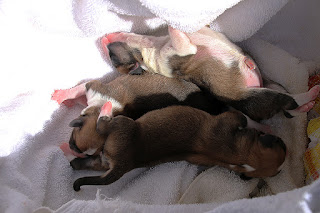
VIDEO shows an emergency Caesarean section for a 2-year-old Corgi.
The Corgi's uterus consists of 2 uterine horns. The left and right uterine horns.
(Image). Normally both uterine horns will be filled with healthy pups in clear water sacs.
In this unusual case,
The entire left uterine horn was filled with black fluid and 3 dead pups and melted placentas.
The entire right uterine horn was filled with normal pups in clear water sacs and placenta.
E:\2017TPV_JUL_SEP\20170716Sun_C_section_mummified_L_horn
Videos
2011 Black vaginal discharge. Breeder phoned Toa Payoh Vets for an emergency Caesarean section.
2012 + 2013 Prepare for surgery. IV drip, isoflurane gas anaesthesia only. No sedatives like propofol IV.
*2014 Left uterine horn. Black fluid filled the inside of the uterus. All 4 pups dead, mummified with no placentas.
Image
Right uterine horn
2016. Right uterine horn is ready for incision
2017, 2018 normal pups pulled out.
Illustration of stitching showing 2 rows
*2019. Uterine horn stitched up in 2 rows of inverting stitches.
lst row is parallel to the incision
*2020 lst row completed. Footage shows 2nd row started.
*2021 end of stitching of 2nd row
2022 End of surgery. Dam cleaned up. Oxytocin, antibiotic and painkiller injections will be given.
2023 + 2024 Healthy pups from right uterine horn.
2029, 2034 Goes back to breeder farm
FOLLOW UP AT BREEDER'S FARM 3 WEEKS LATER.
PUPS OK.
E:\2017TPV_JUL_SEP\20170806Review_3_C_sections
2750 Edit 00:00 - 1:26
Footage shows one of 3 Corgis thriving
Footage shows the single pup (Dachshund) thriving under the poodle. Its mother has not sufficient milk.
The poodle is the surrogate mother of this single pup syndrome Dachshund and one Corgi. The other 2 corgis are nursed by the Corgi dam.
2773 2 Corgi pups
2772 Dachsund single pup syndrome
Pet health and care advices for pet owners and vet students, photography tips, travel stories, advices for young people
Thursday, August 24, 2017
Wednesday, August 23, 2017
3126. Intern Images*****. Be Kind To Pets Veterinary Educational Images - dogs,cats, hamsters, people
https://2010vets.blogspot.com/2018/09/3282-interns-be-kind-to-pets-images-sep.html
DOGS
HAMSTERS
LIZARDS
TURTLES
PEOPLE
https://2010vets.blogspot.com/2018/09/3282-interns-be-kind-to-pets-images-sep.html
Tuesday, August 22, 2017
3125. INTERN VIDEO. Digital evidence of hamster's eye stye inspires confidence in the pet owner
HOOK
Eye problems - painful, blindness unless treated early.
Small creatures like hamsters, eyes too small to be seen by the owner unlilke canine eye problems.
E.g. Pimples in the eyelid known as stye.
BKTP
INTRODUCTION
Why, What, How
Why need vet treatment? Painful, rubbing eye, lose appetite, infections, eyeball rupture
What is stye? Define eye stye
How to convince the owner that her hamster has eye stye?
Digital photography Day 1, Day 7
How is it treated? Eye drops may work. Surgery - incision and drainage under gas anaesthesia in the dwarf hamster.
Hook?
"Your hamster has an eye stye," Vet 2 who was referred to this case by Vet 1 in the branch clinic announced.
The owner of this half-closed, sometimes opened right eyed dwarf hamster applied Vet 2's eyedrops for the next 7 days. The cure was not there. So, the owner sought a 3rd opinion from me.
Nowadays, the young Singaporean pet owner is knowledgeable. Just stating "eye stye" or eyelid pimple in this case is insufficient.

Digital evidence is needed but this takes a lot of time and some skill to develop the evidence to inspire confidence in the young ones. It takes over an hour of digital photoshopping to develop the evidence as shown below.
DAY 1. Gas anaesthesia is necessary to examine and produce sharp photographs the hamster's eye.
Anaesthesia to get proper closed eye examination. Fluorescein eye stain test showed no corneal ulcers. The corneal areas did not show green spots which would have been evidence of eye ulcerations.
I showed the digital evidence of eye stye and advised surgery (incision and drainage) to resolve the problem as the eye pimple is large. The pimple is on the left upper eyelid as at Day 1.
The owner wanted the hamster home on the same day and would apply eye drops and give medication.
DAY 7. The owner returned for the surgery (incision and drainage) after self-treatment for the past 7 days.
The pimples are now on the left and right upper eyelid as at Day 7.
Conclusion
This case shows how digital evidence inspired the owner to return 7 days later for the surgery of incision and drainage to resolve the eye stye problem. The hamster was operated and hospitalised for 3 days.
To acquire digital imaging skills, I practise during my travels and usually daily taking pictures of places and friends and use Photoshop to develop better images as shown below.
--------------------------
Hamster hospitalised for 6 days. On Day 4 after operation, images are taken. They show the conjunctiva of the upper eyelid is inflamed due to the healing of the wound after incising the styes (see Day 7 - 2 styes). Now Day 4 after surgery, no styes on close up image. Show hamster video eye open more, even under operating light (video)
Close up image. Day 4 after incision of the stye.
The styes are not present. The conjunctiva is inflamed as it is early in the healing stage.
Digital photography provides evidence of the scar of the incision (arrows). The wound is healing well.
Conclusion. Many younger Singaporean pet owners are knowledgeable as they research the internet for their pet's diseases.
Digital photography evidence inspire confidence in the owner in the vet. She knows that her pet is properly diagnosed (stye) and treated (post op. Day 4 images).


Day 7 after consultation. The owner came to Toa Payoh Vets for surgery as advised.
This is Day 1 of surgery.

Day 4 of surgery, showing the incision scar (arrow) and the disappearance of the styes.
CREDITS
includes Dr Daniel Sing
FOR MORE INFORMATION
Eye problems - painful, blindness unless treated early.
Small creatures like hamsters, eyes too small to be seen by the owner unlilke canine eye problems.
E.g. Pimples in the eyelid known as stye.
BKTP
INTRODUCTION
Why, What, How
Why need vet treatment? Painful, rubbing eye, lose appetite, infections, eyeball rupture
What is stye? Define eye stye
How to convince the owner that her hamster has eye stye?
Digital photography Day 1, Day 7
How is it treated? Eye drops may work. Surgery - incision and drainage under gas anaesthesia in the dwarf hamster.
Hook?
"Your hamster has an eye stye," Vet 2 who was referred to this case by Vet 1 in the branch clinic announced.
The owner of this half-closed, sometimes opened right eyed dwarf hamster applied Vet 2's eyedrops for the next 7 days. The cure was not there. So, the owner sought a 3rd opinion from me.
Nowadays, the young Singaporean pet owner is knowledgeable. Just stating "eye stye" or eyelid pimple in this case is insufficient.

Digital evidence is needed but this takes a lot of time and some skill to develop the evidence to inspire confidence in the young ones. It takes over an hour of digital photoshopping to develop the evidence as shown below.
DAY 1. Gas anaesthesia is necessary to examine and produce sharp photographs the hamster's eye.
Anaesthesia to get proper closed eye examination. Fluorescein eye stain test showed no corneal ulcers. The corneal areas did not show green spots which would have been evidence of eye ulcerations.
I showed the digital evidence of eye stye and advised surgery (incision and drainage) to resolve the problem as the eye pimple is large. The pimple is on the left upper eyelid as at Day 1.
The owner wanted the hamster home on the same day and would apply eye drops and give medication.
DAY 7. The owner returned for the surgery (incision and drainage) after self-treatment for the past 7 days.
The pimples are now on the left and right upper eyelid as at Day 7.
Conclusion
This case shows how digital evidence inspired the owner to return 7 days later for the surgery of incision and drainage to resolve the eye stye problem. The hamster was operated and hospitalised for 3 days.
To acquire digital imaging skills, I practise during my travels and usually daily taking pictures of places and friends and use Photoshop to develop better images as shown below.
Lens for close up and zoom
--------------------------
Hamster hospitalised for 6 days. On Day 4 after operation, images are taken. They show the conjunctiva of the upper eyelid is inflamed due to the healing of the wound after incising the styes (see Day 7 - 2 styes). Now Day 4 after surgery, no styes on close up image. Show hamster video eye open more, even under operating light (video)
Close up image. Day 4 after incision of the stye.
The styes are not present. The conjunctiva is inflamed as it is early in the healing stage.
Digital photography provides evidence of the scar of the incision (arrows). The wound is healing well.
Conclusion. Many younger Singaporean pet owners are knowledgeable as they research the internet for their pet's diseases.
Digital photography evidence inspire confidence in the owner in the vet. She knows that her pet is properly diagnosed (stye) and treated (post op. Day 4 images).


Day 7 after consultation. The owner came to Toa Payoh Vets for surgery as advised.
This is Day 1 of surgery.

Day 4 of surgery, showing the incision scar (arrow) and the disappearance of the styes.
CREDITS
includes Dr Daniel Sing
FOR MORE INFORMATION
Subscribe to:
Posts (Atom)


