July 19, 2017
Reference: 50 Women's Fashion Icons that changed the world
Lauren Cochrane,
The Design Museum's mission is to celebrate, entertain and inform.
Helps you to wear something different
1. Debbie Harry (1945 - )queen of downtown. Face of punk-pop band Blondie.
Her stage wear uses colour and texture to pack a punch.
Collaborate with designer Stephen Sprouse who was responsible for her asymmetric dresses.
Play with colour including the signature hot pink. Red thigh-high boots and matching red tunic. Peroxide hair and black kohl always present. Still performing. A key reference for young women who want to be a girl in a band and look the part.
2. Tina Chow. Jap mother X American father. Died of AIDS in 1992, aged 41. Lagerfield credited her with minimalism. Concentrate on designing jewellery, furniture and sculpture towards the end of her life. Her cult appeal continues. Photo shows her design - her own bracelets (transparent plastic?),
3. Rei Kawakubo (1942 - ). Her label, Comme des Garcons established in 1969 now at vanguard of fashion. Annual turnover 164 poungs. Dover Street Market dept stores in Tokyo, London and New York. She believes in creating a brand. Cult of her style continues apace.
Wednesday, July 19, 2017
Tuesday, July 18, 2017
Printing Images- what file size do you need
From:
http://www.urban75.org/photos/print.html
http://www.urban75.org/photos/print.html
| Printing images - what file size do you need? A guide to required file sizes for printing out photos [Updated Feb 2016] | |||
|
Image size Pixels
(Virtual Size of Scans) |
Megapixel
rating |
Print size (inches)
at 200ppi |
Print size (inches)
at 300ppi |
|
640 x 480
|
0.3
|
3.2 x 2.4
|
2.1 x 1.6
|
|
1,024 x 768
|
0.8 |
5.1 x 3.8
|
3.4 x 2.5
|
|
1,280 x 960
|
1.2 |
6.4 x 4.8
|
4.2 x 3.2
|
| 1,504 x 1,000 | 1.5 | 7.5 x 5.0 | 5.0 x 3.3 |
|
1,632 x 1,224
|
2.0 | 3.3 x 6.1 | 5.4 x 4.1 |
| 2,000 x 1,312 | 2.6 | 10.0 x 6.6 | 6.7 x 4.4 |
| 2,240 x 1,488 | 3.3 | 11.2 x 7.4 | 7.5 x 5.0 |
| 2,275 x 1,520 | 3.5 | 11.4 x 7.6 | 7.6 x 5.1 |
| 2,272 x 1,704 | 3.9 | 11.4 x 8.5 | 7.6 x 5.7 |
| 2,590 x 1,920 | 5.0 | 13.0 x 9.6 | 8.6 x 6.4 |
|
3,008 x 2,000
|
6.0 |
15.0 x 10.0
|
10.0 x 6.7
|
|
4,256 x 2,848
|
12.1 | 21.3 x 14.2 | 14.2 x 9.5 |
| 4,536 x 3,024 | 13.7 | 22.7 x 15.1 | 15.1 x 10.1 |
| 5,782 x 3,946 | 22.8 | 28.9 x 19.7 | 19.3 x 13.2 |
Professional/online printing Some professional services specify lower resolutions for image sizes, so you could get away with smaller image sizes. Kodak, for example, suggests these resolution/file sizes: For a 4" x 6" print, the image resolution should be 640 x 480 pixels minimum. For a 5" x 7" print, the image resolution should be 1024 x 768 pixels minimum. For an 8" x 10" print, the image resolution should be 1536 x 1024 pixels minimum. For a 16" x 20" print, the image resolution should be 1600 x 1200 pixels minimum. For a 20" x 30" print, the image resolution should be 1600 x 1200 pixels minimum. For a Wallet-size print, the image resolution should be 320 x 240 pixels minimum. Quality vs file size This guide shows you the kind of quality you can expect when you're printing out your photos. | |||
|
Digital camera resolution vs print quality
|
||||||||
|
Capture Resolution
|
Video Display
|
Print Size
|
||||||
|
2x3"
|
4x5"/4x6"
|
5x7"
|
8x10"
|
11x14"
|
16x20"
|
20x30"
|
||
|
320x240
|
OK
|
Good
|
OK
|
Crap
|
Crap
|
Crap
|
Crap
|
Crap
|
|
640x480
0.3Megapixel |
Good
|
Excellent
|
Good
|
Crap
|
Crap
|
Crap
|
Crap
|
Crap
|
|
800x600
|
Excellent
|
Photo Quality
|
Very Good
|
Reasonable
|
Crap
|
Crap
|
Crap
|
Crap
|
|
1024x768
|
Excellent
|
Photo Quality
|
Excellent
|
Good
|
OK
|
Crap
|
Crap
|
Crap
|
|
1280x960
1 MP |
Excellent
|
Photo Quality
|
Photo Quality
|
Very Good
|
Good
|
Crap
|
Crap
|
Crap
|
|
1536x1180
|
Excellent
|
Photo Quality
|
Photo Quality
|
Excellent
|
Very Good
|
OK
|
Crap
|
Crap
|
|
1600x1200
2MP |
Excellent
|
Photo Quality
|
Photo Quality
|
Photo Quality
|
Very Good
|
OK
|
OK
|
Crap
|
|
2048x1536
3 Megapixel |
Excellent
|
Photo Quality
|
Photo Quality
|
Photo Quality
|
Excellent
|
Good
|
OK
|
OK
|
|
2240x1680
4 Megapixel |
Excellent
|
Photo Quality
|
Photo Quality
|
Photo Quality
|
Photo Quality
|
Very Good
|
Good
|
OK
|
|
2560x1920
5 Megapixel |
Excellent
|
Photo Quality
|
Photo Quality
|
Photo Quality
|
Photo Quality
|
Excellent
|
Very Good
|
Very Good
|
|
3032x2008
6 Megapixel |
Excellent
|
Photo Quality
|
Photo Quality
|
Photo Quality
|
Photo Quality
|
Photo Quality
|
Excellent
|
Very Good
|
3072x2304
7 Megapixel |
Excellent
|
Photo Quality
|
Photo Quality
|
Photo Quality
|
Photo Quality
|
Photo Quality
|
Excellent
|
Excellent
|
3264x2448
8 Megapixel |
Excellent
|
Photo Quality
|
Photo Quality
|
Photo Quality
|
Photo Quality
|
Photo Quality
|
Photo Quality
|
Excellent
|
10 Megapixel +
|
Excellent
|
Photo Quality
|
Photo Quality
|
Photo Quality
|
Photo Quality
|
Photo Quality
|
Photo Quality
|
Photo Quality
|
| Crap | Horribly pixelated blocky mess, like something out of 1987 |
| OK | Looks reasonable enough, but nowhere near photo quality |
| Good | Pretty good all round, with a fair bit of detail |
| Very Good | This will be fine for most uses, unless you get up reeeeal close |
| Excellent | As good as a photo for most purposes |
| Photo Quality | Woohoo! Fantastic quality print, full of detail
and perfect for your picture frame or for your showing off your very
best shots. |
Sunday, July 16, 2017
3100. A 16-year-old Maltese has a large left perineal hernia recurred some 7 years after repair.
Another case of a gigantic perineal hernia. The owners had consulted Vet 3 some months ago and was referred to a vet specialist who would be more costly. So a friend referred them to me.
Some 7 years ago, Vet 1 had operated but the hernia recurred some months later. Vet 1 referred to Vet 2 who operated but the hernia recurred. So Vet 2 did muscle transposition surgery,cystopexy and colopexy. This last operation lasted around 6 years. The dog was neutered young and this is a rare case as 99% of the perineal hernias in male dogs are not neutered. The owners did not want to go back to Vet 2 for some reasons although his fees were reasonable at around $1,500 7 years ago.
July 2017.
"Your dog is very old, at 16 years and has heart disease," I advised against any surgery to repair this hernia. "She can pee. She eats like a horse."
"But she cannot poop for the past 5 days," the mother and adult daughter said. "Her swelling is very painful too."
"She needs to be anaesthesized for me to evacuate the stools manually," I said there would be anaesthetic risks but the owner had no choice. She signed the consent form. Anaesthesia would be short as I used fingers to extract the semi-hard stools from the rectum. Dog was OK and stayed one day inpatient.
X RAYS


NO SURGERY
"Your dog is not fit for surgery as he has heart disease," I said. "Surgery using mesh implant will take around one hour. He can live another 2 years or more if his heart disease is controlled by medication."
1. Reduce the swelling 6 times a day so that the rectum does not get trapped.
2. Give lactulose daily for 7 days, then alternate days for week 2 and 3.
3. Switch to Prescription Diet W/D as it has higher fibre content. If W/D works, no need to give lactulose.
4. Heart disease medication daily for 2 weeks first and review.
FOLLOW UP
1. Much less coughing after heart medication is given
2. Oral 3 ml lactulose per day. Passed stools, not watery.
3. Swelling is there, but the dog is not constipated as before evacuation.
TIPS
The hernia had recurred some 6 years after the 2nd surgery but the dog was able to poop.
The vet refused to prescribe more lactulose without examination and so the dog got constipated after a while.
3099. An unusual Sunday emergency caesarean section - mummified foetuses.
Sunday Jul 16, 2017
Emergency C-section phone call at 9 am. 2-year-old Corgi, 61st day pregnant, lst litter, passed out viscous blackish vaginal discharge.
So, rushed down to clinic. Breeder was there.
"This Corgi is the daughter of the Corgi that died after C-section by Vet 1," the Myanmar assistant told me. "This is her first litter. She was active yesterday, but today, passed out black vaginal discharge." Breeders never forget any death after C-section and would avoid the vet. As for me, I had no deaths so far.
Unusual C-section.
The left uterine horn was filled with viscous black liquid enveloping 4 below-normal sized puppies. All 4 puppies were mummified and dead. One was very small in size. The right uterine horn had 3 well-formed pups but the placentas had "melted" and so the pups detach easily from the uterus. I gave them to the breeder who is hands-on and prefers to revive the pups himself as he said he is most experienced. Some vets would not permit him to do so.
Only isoflurane gas using cone first, before size 7.5 endotracheal tube intubation.
TIPS
The dam fought against cone anaesthesia vigorously. This is expected. "Take off the cone when the dam struggles, " I said to my assistant Judy. "Sometimes, the dam is choked. Let her breathe without the cone and put in again. She did this. The dam was not struggling and became anaesthesized soon.
At my 2nd last stitch, the dam appeared to "choke". I said, "The endotracheal tube may be irritating the trachea, pull it out a bit." It is hard to know what causes this behaviour as she was on smooth anaesthesia. I stitched up the last stitch and got the tube out.
3 live vigorous crying puppies. The breeder saw the mummified puppies and was satisfied with having 3 live ones. As to the cause of 4 mummified puppies in one uterine horn, it is a mystery. In any case, the 3 normal puppies would have died if the breeder proscratinate another hour as the placentas had detached and melted especially for 2 of them. See video.
Friday, July 14, 2017
3098. A 9-year-old Maltese has a gigantic perineal hernia. Mesh implant.


Thursday, July 13, 2017
3097. A male dwarf hamster urine marks causing itchy lower body.
July 13, 2017
PUDDING, male, dwarf hamster, born June 2016.
Today, the young man consulted me and said: "My hamster still has an itchy armpit after you had operated on her left elbow wart 5 weeks ago!" I was surprised and got my assistant to video the new medical condition.
"5 weeks ago, I removed the left elbow wart which was 2 mm across," I said. "There was no elbow problem."
The hamster squealed twice as I examined the sore right armpit using a small torch-light.
"It is the right armpit," my assistant pointed out.. "The wart was on the left elbow!"
A rough red inflamed patch of loose skin around 4mm x 4mm was present on the right elbow. So this was a new condition.
HISTORY
Dec 28, 2016
Lives alone. Overweight 63 g. Urine spraying on exercise wheel, food bowl, corner and litter. Sleeps on urine.
1. URINE SCALDING. Whole lower body has no hair. Inflamed and itchy. Diagnosed: Ventral dermatitis. Advised neuter.
2. Left ear wart 4mm x 4 mm. Electro-excised ear wart.
Jan 17, 2017
63g
Lethargic
Scratches both ears, flanks and bites paws. Hair loss. Paws inflamed.
Ventral and flank dermatitis.
Otitis externa both ears.
Open vertical ear canals, irrigate
Clip bald under anaesthesia
Cleaning scent gland
Jun 8, 2017
68g
Left elbow wart 4 x3 mm across electro-excised.
No bleeding.
Jul 13, 2017
69g
Right armpit skin inflamed and biting the area. 5 mm x 5 mm.
CONCLUSION
Owner did not neuter as the hamster no longer urine mark.
Did not manage to reduce weight. No more sand bath.
Tuesday, July 11, 2017
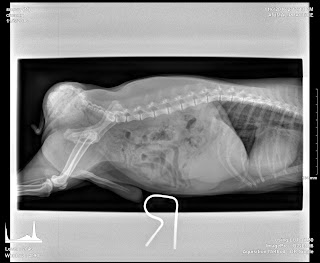
3096. 16-year-old Cocker Spaniel had swollen abdomen
Blood test showed liver disease.
X-ray showed liver tumours from mammary gland tumours.
The dog had a swollen abdomen due to liver tumours. Passed away today.


The dog was much loved.
X-ray showed liver tumours from mammary gland tumours.
The dog had a swollen abdomen due to liver tumours. Passed away today.


The dog was much loved.
Subscribe to:
Posts (Atom)