Sunday 26, 2012. Bright sunshine. Went to NATAS fair at Changi expo at 9.30 am to see what was available in the travel industry and new ideas on marketing. My assistant phoned to say I had an appointment at 11 am.
"Sheltie very itchy below the penis," said the gentleman. I thought it was penile trauma or inflammation of the prepuce. On seeing the dog with Dr Daniel, I saw the red circular spots and the crusted centre. "Ringworm," I said. "Had the dog been to the groomer recently?"
"2 weeks ago," the owner said.
I showed the ringworm hairs to Dr Daniel. He was not convinced saying that those could be damaged hairs, due to the dog licking the ventral area, causing inflammation and rashes.
He didn't buy into my microscopic exam of ringworm hair. Each vet has his own ideas but there are certain presentation that cannot be ignored.
"Circular rings, reddish with crusty scales in the centre," i said. These are typical presentation.
"Needs confirmation with fungal culture," he said. This is what the professors teach.
"Yes," I said. "But the owner cannot wait 2 weeks for the culture to prove it is ringworm. He wants prompt action and successful outcome."
In fact the couple had a bare patch on the dog's left shoulder area diagnosed "ringworm" by Vet 1 from Toa Payoh Vets. Since it did not resolve and the dog was still scratching, he went to Vet 2 who diagnosed demodectic mange.
"How many mites he showed you?" I asked the gentleman banker.
"One mite," he described the cigar shape.
"Well, it is possible that it is demodectic mange on that hot spot," I said. "In any case, demodectic mites are normally present in dogs, as are bacteria in our mouth. Ringworm takes more than 1 week to cure."
I did not want to defend Vet 1's "misdiagnosis" as the gentleman would not believe me. Vet 2 had cured his dog and that was results. "Yellow pimples," he said.
Wednesday, February 29, 2012
888. Megaesophagus in the older dog - treatment
Regurgitation is the presenting sign. Mistaken for "vomiting" which is a projectile action.
D/D for regurgitation
1. Esophageal obstruction. Vascular ring anomaly, stricture, foreign body, neoplasia (spirocerca lupi).
2. Motility disorders. Megaoesophagus - congential
- acquired --- primary (idiopathic)
--- secondary
Dysautonomia
3. Inflammatory. Esophagitis (secondary to protracted vomiting), caustic irriation. Hiatal hernia. Gastro-esophageal reflux.
4. Others. Diverticula, Broncho-esophageal fistula.
5. Idiopathic.
DIAGNOSIS
1. History - Other clinical signs, young or older breed, large breed
2. Physical exam - incl full neurological exam, thorough thoracic auscultation (aspsiration pneumonia, caudal lobes on X-ray)>
THORACIC X-RAY. Needs 3 views. 2 lateral + V/D. Look for aspiration pneumonia, cranial mediastinal mass, air in esophagus (one lateral without sedation as drugs can cause air in esophagus).
Contrast studies. Liquid barium using plain X-rays, evidence of strictures, vascular ring anomaly (puppies).
Food and barium dynamic studies (fluoroscopy) to assess motility of esophagus.
Other investigations to identify underlying problems.
TREATMENT depends on causes
Distemper, autonomic drugs, toxicity (lead, thallium, anti-cholilnesterase), esophigitis, systemic lupus erythematosus, hypoadrenocorticisum, thyoma, myasthenia gravis (focal, most common cause, auto-antibodies produced againt Ach receptors), polymyositis.
1. Vertical feeding - Feed from a height. Bailey's chair. 1 hour upright feeding.
2. Bethanechol or cisapride for myasthenia gravis
CASE STUDY
Use of barium meal showed esophagus enlarged in two areas, convincing the owner of the diagnosis. Plain X-ray not so clear in showing megaesophagus.
2. Dietary - canned food, meat balls. No dry food.
3. If myasthenia gravis, require anit-cholinesterase drugs and immune suppression.
bethanechol or cisapride
Tuesday, February 28, 2012
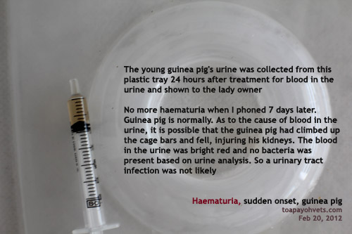
Follow up on guinea pig that suddenly passed blood in urine
Guinea pig, male, young, had passed blood in the urine some weeks ago. Vet 1 diagnosed UTI. Recovered after some days.
Again blood in urine. Sent to Toa Payoh Vets. Consulted me with Dr Daniel. Another recurrence of UTI? "The blood looks clean and bright red," I said. "It is unlikely to be UTI. Collect some urine for urine analysis."
How to collect urine? This is not a male dog or cat. A small male guinea pig that is much loved by his mistress who had consented to more detailed investigations rather than the usual injection and go home to wait and see. "Practise evidence-based medicine," I always advise my vets. "Feeling that it is not UTI or it is UTI is not sufficient. Feeling is not not enough". So, I had to practise what I preach and collect the urine from this little one.
Well, one must improvise as the urine must be clean and not be contaminated from faecal stools. Pictures showed.


After one day of treatment and rest, the guinea pig went home. I phoned the owner yesterday, Feb 27, 2012, around 7 days later. No more problem since going home.
FOLLOW UP ON A LITTLE GUINEA PIG'S HEALTH
Her reply on Feb 27, 2012 is as follows:
Hi Dr Sing,
The only time he's elevated to a height is when I carry him to bathe once a week. However, I have never dropped him before. He doesn't struggle at all. No one else carries him, they usually pat him when he's walking around on the floor (with supervision). When I carry him, I am seated on e floor too.
His whole cage is cleaned and wiped down with diluted vinegar or pet surface sanitizer (by byopet) at least twice a day
(morning and evening) or more.
After he got back from your clinic, he's been eating well n drinking well, a lot of hay and pellets. I feed him small portions of vegetables twice a day (either lettuce, baby carrots or cucumbers).
Thank you.
Sent from my iPhone
Again blood in urine. Sent to Toa Payoh Vets. Consulted me with Dr Daniel. Another recurrence of UTI? "The blood looks clean and bright red," I said. "It is unlikely to be UTI. Collect some urine for urine analysis."
How to collect urine? This is not a male dog or cat. A small male guinea pig that is much loved by his mistress who had consented to more detailed investigations rather than the usual injection and go home to wait and see. "Practise evidence-based medicine," I always advise my vets. "Feeling that it is not UTI or it is UTI is not sufficient. Feeling is not not enough". So, I had to practise what I preach and collect the urine from this little one.
Well, one must improvise as the urine must be clean and not be contaminated from faecal stools. Pictures showed.
After one day of treatment and rest, the guinea pig went home. I phoned the owner yesterday, Feb 27, 2012, around 7 days later. No more problem since going home.
FOLLOW UP ON A LITTLE GUINEA PIG'S HEALTH
Her reply on Feb 27, 2012 is as follows:
Hi Dr Sing,
The only time he's elevated to a height is when I carry him to bathe once a week. However, I have never dropped him before. He doesn't struggle at all. No one else carries him, they usually pat him when he's walking around on the floor (with supervision). When I carry him, I am seated on e floor too.
His whole cage is cleaned and wiped down with diluted vinegar or pet surface sanitizer (by byopet) at least twice a day
(morning and evening) or more.
After he got back from your clinic, he's been eating well n drinking well, a lot of hay and pellets. I feed him small portions of vegetables twice a day (either lettuce, baby carrots or cucumbers).
Thank you.
Sent from my iPhone
886. Closed pyometra email
E-MAIL TO DR SING DATED FEB 28, 2012
Hi Dr Sing
I would appreciate your advice on my dog's case.
My dog is an unspayed female Shih Tzu whose age is estimated to be around 5-6yr old(no exact age as we adopted her when she was found abandoned and in bad condition). Her weight is 3.95kg.
Her last heat ended around 30th November 2011.
She is suffering from bad case of skin problem and is drinking more water and has slimmed down. Her appetite is good with no vomiting and diarrhoea. No discharge or blood in urine observed.
I sent my dog to the vet for her skin problem and the blood test results showed elevated WBC (26.92+). As she is observed to be drinking more water coupled with elevated WBC and her unspayed status, the vet suspected it might be closed pyometra.
No X-ray or ultrasound was done due to financial concerns. Skin scrape was negative for demodex. According to the vet, blood work showed no other abnormalities. However, her T4 readings is quite low (1.3) on the normal range (1.1-4.0 UG/DL).
I can email you the blood work results once I get them scanned should you require them.
Vet prescribed Baytril 50mg tablet(1/2 tablet daily) and to recheck in 2 weeks time. She is also too skinny and vet gave Troy Nutripet gel to supplement her diet.
I am very concerned about the dangers of pyometra and would like to seek your advice. As her body is already stressed by her skin condition and her malnourished state, I am very lost.
I do not know if putting her on the antibiotics and supplementing her health in preparation for the surgery while observing for sudden changes before the next consult would be advisable? Or should I just opt for early surgery since her bloodwork suggests generally healthy organ functions.
Do you think it is possible for her to be operated by you without performing the imaging tests? What is your estimated total charges for the surgery? Please kindly advise. Thank you very much.
Best Regards
Name given
E-MAIL REPLY FROM DR SING DATED FEB 28, 2012
E-MAIL REPLY FROM DR SING DATED FEB 28, 2012
I am Dr Sing. Thank you for your email.
It is risky to diagnose closed pyometra by email without examination of the patient. I will give you my opinions based on your email report on the understanding that it is best to have an examination by me, if practical.
1. Closed pyometra occurs in a dog that has her heat period around 2-3 months ago and your dog was in this situation. She had increased white cell count and was drinking more. I presume she has weight loss over the last 2-3 months. Pl email your vet's reports and blood test results.
2. However, skin disease and poor health can also cause increased white cell count and thirst due to skin infections and itchiness. Drugs may also increase thirst or appetite if your vet had earlier treated the dog.
3. As your dog has good appetite and no vomiting, it is possible that, at the time of consultation, she does not have closed pyometra, assuming she had not been given any drug to treat her skin disease or other health problems.
QUESTIONS
3.1. Did your vet palpate the abdomen and let you know the results of his or her palpation as to whether he or she felt an enlarged uterus? Abdominal palpation is an alternative to X-rays and ultrasound and if the uterus is enlarged, it can be palpated by the vet. Closed pyometra can be confirmed just by palpation but imaging is sometimes necessary to "convince" the owner of the presence of the enlarged uterus.
3.2 Did your dog have fever at the time of consultation? What was the rectal temperature?
3.3 When did the weight loss start? Was it after November 30, 2011 or was the dog losing weight for many months?
4. In reply to your questions about spaying your dog without imaging, it is possible. My estimated charges for the operation would be around $250-$300 for a normal spay and around $500 - $600 for a pyometra spay as this takes a longer time and requires IV drip.
5. It is prudent to wait 2 weeks or longer for your dog to recover from the infection and put on weight, before any spay surgery is attempted. The skin disease should be cured first. The cause of the weight loss may be due to the start of closed pyometra if weight loss is recent.
Another blood test can be done to check the total white cell count prior to surgery, if possible.
6. Closed pyometra is sometimes difficult to diagnose when the uterus is not full of pus. Usually there is loss of appetite. Yet you said your dog has "good" appetite but is losing weight. Why? A typical Shih Tzu with good appetite at middle age should be weighing more than 3.95 kg, at around 5-6kg. Therefore, I don't think that your dog has "good appetite."
I hope the above answers your questions.
Saturday, February 25, 2012
885. Veterinary Surgery Audit at Toa Payoh Vets by Dr Sing Kong Yuen
There is a need to be efficient and productive when vets operate at Toa Payoh Vets.
1. Name of file - 2012TPV Anaesthesia Record
TP Reference - 42373
Date of surgery - 24.2.12
Name of owner/representative: Mr ...
2. Patient
Name:
Age: 15 months
Weight: 5.8 kg
Temp: 38.6C
Microchip No.
3. Surgery
Procedure: Spay. Vet: Dr Sing Kong Yuen
Suture type: 2-0 Polysorb (Braided lactomer, Cutting 3/8 24 mm)
Suture packets used: One packet
Spay certificate No: 1675
4. Anaesthesia
Induction Drugs: Domitor + Ketamine IV
Dose calculation
Weight (kg)
Age
Dom
Ket
10
Young
0.4
0.5
5
Young
0.23
0.29
Given 50 %
0.1
0.15
One syringe with 0.25 ml. I added 0.15 ml normal saline = 0.4 ml IV
Top up
Isoflurane + O2 at 0.5-1
% for 21 minutes (C-B)
Isoflurane graph charting % at 5-minute intervals
Summary:
Isoflurane maintained at: 0.5 - 1.0% for 21 minutes
Route: IV
A. Time of injection of induction drug: 5.07pm
B. Time of isoflurane gas first given: 5.11pm
C. Time of isoflurane gas stopped: 5.32pm
D. Time of first skin incision: 5.17pm
E. Time of completion of skin stitching: 5.33pm
E-A = 26 minutes
E-D = 16 minutes
C-B = 21 minutes
MY AUDIT OF VETERINARY SURGERY IS BASED ON:
SURGICAL TIME TO SPAY A DOG IN THIS CASE
It took 16 minutes to spay this dog (E-D). I took 2 attempts to hook left the ovary out and had two ligatures (one transfixing and one normal) done on the uterine body. If I hook the ovary at the first attempt and ligate the uterine body once, the timing should be around 13 minutes. No bleeding seen in this case.
TIPS
The skin incision was around 1.8 cm long and the location was right, around 2 cm from the umbilical scar. The 4 legs were stretched very taut as I find this procedure much easier to hook out the left ovary. I used the scalpel to cut the ovarian ligament instead of fingers to snap it. Then I ligated the ovarian ligament area once. The dog's head was on my left. I raised the hydraulic table to suit my height as with too low the table height, I had to bend down and this would delay surgery. No swab was needed in this case. 2/0 absorbable suture x 1 packet was used. I seldom see the owner coming back for stitch removal as the stitches dissolve in 14-28 days. This saves the owner one trip and time.
5. OTHER MATTERS
Spay timing:
Skin incision: 5.17pm
Linea alba incision: 5.17pm
Left ovary hooked out: 5.19pm
Left ovarian ligament incised: 5.19pm
Left ovary clamped: 5.20pm
Left ovary ligated: 5.21pm.
Right ovary hooked out: 5.21pm
Right ovarian ligament incised: 5.22pm
Right ovary clamped: 5.22pm
Right ovary ligated: 5.23pm
Uterine body (UB) clamped: 5.24pm
UB transfixation ligature right side: 5.25pm
UB transfixation ligature left side: 5.26pm
UB 2nd ligation round UB: 5.27pm
UB incised: 5.29pm
UB checked for bleeding before putting into abdomen: 5.29pm
Linea alba stitched: 2 simple interrupted sutures: 5.29pm
Finger palpate linea alba (2 cm) for hole: 5.31pm.
Skin stitched: 5.31pm. First horizontal mattress
Isoflurane gas stopped: 5.32pm
Skin stiched: 5.32 pm. Second horizontal mattress
Uterus weight: 26 g. Owner said, not pregnant before
Spay certificate No. 1675
Dog's surgical wound bandaged, given baytril 0.6 ml and tolfedine 0.6 ml SC
E-collar. Home Baytril 50 mg x 2 (1/2 tab sid), Tolfedine 6 mg x 16 tab (4 tab sid)
6.30 pm phoned owner. Take dog home. Awake.
Website is at:
http://www.sinpets.com/F5/20120233SPAY-audit-surgical-anaesthetic-time-Singapore_ToaPayohVets.htm
Friday, February 24, 2012
884. Anaesthetic & Surgical Record for Toa Payoh Vets
The following is to monitor the productivity of veterinary anaesthesia and surgery and for instructions and review
1. Name of file
TP Reference
Date of surgery
2. Patient
Name
Age
Weight
Temperature
3. Surgery
Procedure
Vet
Spay/Neuter Certifcate No:
4. Anaesthesia
Induction Drugs
Dose calculation
Route
Isoflurane graph charting % at 5-minute intervals
Review: Isoflurane maintained at: 0.5 - 1.0% for 21 minutes
A. Time of injection of induction drug
B. Time of isoflurane gas first given
C. Time of isoflurane gas stopped
D. Time of first skin incision
E. Time of completion of skin stitching
E-A = minutes
E-D = minutes
C-B = minutes
5. OTHER MATTERS
Vet signature:
Using dog spayed by Dr Sing as an example
1. Name of file - 2012TPV Anaesthesia Record
TP Reference - 42373
Date of surgery - 24.2.12
2. Patient
Name: Jo Jo
Age: 15 months
Weight: 5.8 kg
Temp: 38.6C
3. Surgery
Procedure: Spay
Vet: Dr Sing Kong Yuen
Suture type: 2-0 Polysorb (Braided lactomer, Cutting 3/8 24 mm
Suture packets used: One packet
Spay certificate No: 1675
4. Anaesthesia
Induction Drugs: Domitor + Ketamine IV
Dose calculation
Wt Age D K
10 young 0.4ml 0.5ml
5.8 young 0.23 0.29
@50% 0.1 0.15 given = 0.26 ml. Add 0.14 normal saline = 0.4 ml IV
Isoflurane graph charting % at 5-minute intervals
Isoflurane maintained at: 0.5 - 1.0% for 21 minutes
Route: IV
A. Time of injection of induction drug: 5.07pm
B. Time of isoflurane gas first given: 5.11pm
C. Time of isoflurane gas stopped: 5.32pm
D. Time of first skin incision: 5.17pm
E. Time of completion of skin stitching: 5.33pm
E-A = 26 minutes
E-D = 16 minutes
C-B = 21 minutes
5. OTHER MATTERS
Spay timing:
Skin incision: 5.17pm
Linea alba incision: 5.17pm
Left ovary hooked out: 5.19pm
Left ovarian ligament incised: 5.19pm
Left ovary clamped: 5.20pm
Left ovary ligated: 5.21pm.
Right ovary hooked out: 5.21pm
Right ovarian ligament incised: 5.22pm
Right ovary clamped: 5.22pm
Right ovary ligated: 5.23pm
Uterine body (UB) clamped: 5.24pm
UB transfixation ligature right side: 5.25pm
UB transfixation ligature left side: 5.26pm
UB 2nd ligation round UB: 5.27pm
UB incised: 5.29pm
UB checked for bleeding before putting into abdomen: 5.29pm
Linea alba stitched: 2 simple interrupted sutures: 5.29pm
Finger palpate linea alba (2 cm) for hole: 5.31pm.
Skin stitched: 5.31pm. First horizontal mattress
Isoflurane gas stopped: 5.32pm
Skin stiched: 5.32 pm. Second horizontal mattress
Uterus weight: 26 g. Owner said, not pregnant before
Spay certificate No. 1675
Dog's surgical wound bandaged, given baytril and tolfedine SC
E-collar. Home Baytril 50 mg x 2 (1/2 tab sid), Tolfedine 6 mg x 16 tab (4 tab sid)
6.30 pm phoned owner. Take dog home. Awake.
Vet signature:
1. Name of file
TP Reference
Date of surgery
2. Patient
Name
Age
Weight
Temperature
3. Surgery
Procedure
Vet
Spay/Neuter Certifcate No:
4. Anaesthesia
Induction Drugs
Dose calculation
Route
Isoflurane graph charting % at 5-minute intervals
Review: Isoflurane maintained at: 0.5 - 1.0% for 21 minutes
A. Time of injection of induction drug
B. Time of isoflurane gas first given
C. Time of isoflurane gas stopped
D. Time of first skin incision
E. Time of completion of skin stitching
E-A = minutes
E-D = minutes
C-B = minutes
5. OTHER MATTERS
Vet signature:
1. Name of file - 2012TPV Anaesthesia Record
TP Reference - 42373
Date of surgery - 24.2.12
2. Patient
Name: Jo Jo
Age: 15 months
Weight: 5.8 kg
Temp: 38.6C
3. Surgery
Procedure: Spay
Vet: Dr Sing Kong Yuen
Suture type: 2-0 Polysorb (Braided lactomer, Cutting 3/8 24 mm
Suture packets used: One packet
Spay certificate No: 1675
4. Anaesthesia
Induction Drugs: Domitor + Ketamine IV
Dose calculation
Wt Age D K
10 young 0.4ml 0.5ml
5.8 young 0.23 0.29
@50% 0.1 0.15 given = 0.26 ml. Add 0.14 normal saline = 0.4 ml IV
Isoflurane graph charting % at 5-minute intervals
Isoflurane maintained at: 0.5 - 1.0% for 21 minutes
Route: IV
A. Time of injection of induction drug: 5.07pm
B. Time of isoflurane gas first given: 5.11pm
C. Time of isoflurane gas stopped: 5.32pm
D. Time of first skin incision: 5.17pm
E. Time of completion of skin stitching: 5.33pm
E-A = 26 minutes
E-D = 16 minutes
C-B = 21 minutes
5. OTHER MATTERS
Spay timing:
Skin incision: 5.17pm
Linea alba incision: 5.17pm
Left ovary hooked out: 5.19pm
Left ovarian ligament incised: 5.19pm
Left ovary clamped: 5.20pm
Left ovary ligated: 5.21pm.
Right ovary hooked out: 5.21pm
Right ovarian ligament incised: 5.22pm
Right ovary clamped: 5.22pm
Right ovary ligated: 5.23pm
Uterine body (UB) clamped: 5.24pm
UB transfixation ligature right side: 5.25pm
UB transfixation ligature left side: 5.26pm
UB 2nd ligation round UB: 5.27pm
UB incised: 5.29pm
UB checked for bleeding before putting into abdomen: 5.29pm
Linea alba stitched: 2 simple interrupted sutures: 5.29pm
Finger palpate linea alba (2 cm) for hole: 5.31pm.
Skin stitched: 5.31pm. First horizontal mattress
Isoflurane gas stopped: 5.32pm
Skin stiched: 5.32 pm. Second horizontal mattress
Uterus weight: 26 g. Owner said, not pregnant before
Spay certificate No. 1675
Dog's surgical wound bandaged, given baytril and tolfedine SC
E-collar. Home Baytril 50 mg x 2 (1/2 tab sid), Tolfedine 6 mg x 16 tab (4 tab sid)
6.30 pm phoned owner. Take dog home. Awake.
Vet signature:
Thursday, February 23, 2012
883. Lateral saphenous artery - Golden Retriever tumour in front of knee
The case of the old Golden Retriever with a large knee tumour.
"Your vet quoted $300 for the operation," the mother said.
"Who was the vet?" I asked as the surgery to excise this large knee tumour will take a long time, at least one hour. I had quoted $500. The owner did not want blood test or histopathology of the tumour and that would save some money. Still, $500 for the whole procedure including drugs, e-collar and post-op care was very low.
ANAESTHESIA
50% of domitor + ketamine IV according to my guidelines written previously.
The dog was old and at 50% was really knocked out. A whiff of the isoflurane gas + O2 after 5 minutes of sedation enabled intubation.
SURGERY
I demonstrated the surgery to Dr Daniel by operating together as this would not be a simple surgery as removing a tumour from the side of the body where there is a lot of skin. Here, the tumour was massive at 7 cm x 8 cm x 5 cm and if the textbook advice is to be followed, a wide resection meant insufficient skin for stitching. A wide resection is important to remove all tumour cells but an big knee wound due to insufficient skin area to close is deadly for the dog post-op as bacterial infection comes in over time.
Use marker pen to know how to excise.
BLEEDING ARTERY
There is one spurting artery of around 2 mm in diameter from the skin surface lateral to the tumour. "It is the lateral saphenous artery," Dr Daniel said. I advised a "purse-string" suture with the 2/0 absorbable and he did it. The bleeding stopped. But profuse bleeding from all other tissues continued. Swab, swab, swab, swab.
"A bi-polar electrode will be most useful," he said.
"In old dogs, the faster the surgery is done, the safer it is for this 8-year-old. I excised the tumour fast and started stitching. The bleeding continue unabated as there were numerous veins and smaller arteries. In theory, the bi-polar electrode would be used to coagulate. I could also use the coagulation electrode by switching to it from excision electrode."
"Look, the tongue and gum colour of the Golden Retriever is getting purplish," I said. In theory, it is best to stop all bleeding. This prolonged anaesthesia and the dog dies.
In practice, I stitched up the wound and used bandaging. A live patient is what the owner wants, not a clean no-bleeding surgical wound. This is the good outcome that is what text books don't teach.
You can see pictures at www.toapayohvets.com now. Will update again.
Myanmar tourism has exploded as at Feb 2012
On Feb 22, 2012, I spoke with my Myanmar travel agent partner about the latest in Myanmar tourism since the U.S has made contacts with the politicians and Aung San Sui Kyi is allowed to be in politics openly.
The following are the latest happenings in Myanmar tourism.
1. A sudden surge in tourist arrivals
2. Very difficult to find 4- and 5-star accommodation in Yangon's hotels due to the increase in businessman visitors from the Middle East and from Non-Governmental Organisations. The Middle East tourists rent the whole floor and pays 50% more, according to my partner.
3. Therefore, there is insufficient hotel rooms for tourists who want 4- and 5-star hotel. There are 2- and 3-star accommodation and they are safe, decent and clean too.
4. The administrators want to build a subway in Yangon and had approached the governments of Singapore and Japan as both have the experience.
The investors are given the land to build and both share the profits.
Tuesday, February 21, 2012
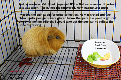
881. Blood in the guinea pig's urine again. Why?
"UTI" the owner said the previous vet had diagnosed UTI when her guinea pig had passed blood in the urine.
No urine test was taken by the first vet. She wanted another opinion from me.
"UTI is common," I said. "I need to ward it and do a urine collection to check the urine."
How to collect urine from a guinea pig? I remembered my sheep metabolic studies lectures in Animal Nutrition in the 3rd year. That was 4 decades ago. Improvise. I looked for a clean pan, not the usual pee pan. I found two. See images.


Will update later.
Tentative diagnosis is traumatic injury as the guinea pig still has good appetite and has not passed blood in the urine on 2nd day of hospitalisation after being treated with medication.
Image from link from toapayoh vets as blogger.com seems to have some problems in layout when images are posted directly to it.
Monday, February 20, 2012
880. Sunday's interesting cases at Toa Payoh Vets
Feb 19, 2012
Bright sunshine day
CASE 1.
"My guinea pig has UTI. He passes blood in his urine," the young lady's GP carrier had two large spots of blood spread onto the white tissue paper. "He had passed blood before and recovered after antibiotics," she had the GP treated by 2 vets elsewhere a few weeks ago. The GP of one year old had excellent appetite and was bright.
"Was urine collected for analysis?" I asked as I shared the case with Dr Daniel. Seldom do vets collect urine from a GP. It is just not the thing to do. I mean, how do you collect urine from a 500g GP? Male dogs can be catheterised to collect urine. But a GP?
"No," she said.
"A urine analysis is most important," I said. "I need to ward the GP for 2 days to observe and collect urine."
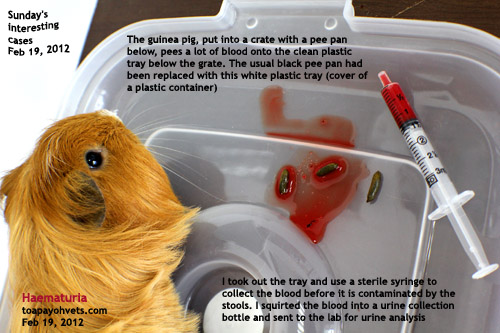
How to do it? I used two clean plastic tray covers under the grated floor. See image.
Fresh red blood. Bright red as if there was some bleeding internally inside the bladder. Or kidney? More passed. I wondered if the GP would bleed to death.
1. UTI? That's the primary diagnosis. The other vets had diagnosed UTI and so the owner assumed UTI.
2. Certain types of food causing reddish blood. But this was pure red blood.
3. Coagulopathy? Dr Daniel suggested.
The GP passed more than 10 ml of blood. I collected 1.5 ml from the tray and sent to the lab.
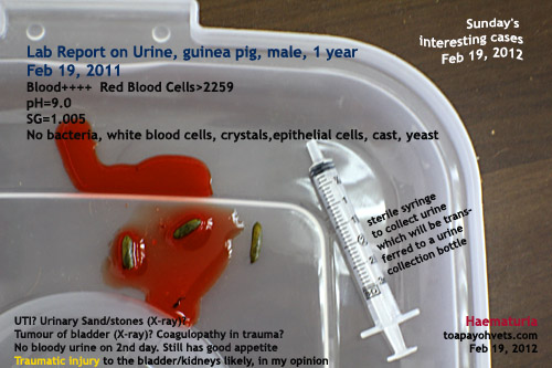
URINE ANALYSIS
Blood+++. pH=9.0. Negative for bacteria, crystals, WBC.
The GP did not pass blood after 24 hours. He ate only fresh apples and vegetables.
Bright sunshine day
CASE 1.
"My guinea pig has UTI. He passes blood in his urine," the young lady's GP carrier had two large spots of blood spread onto the white tissue paper. "He had passed blood before and recovered after antibiotics," she had the GP treated by 2 vets elsewhere a few weeks ago. The GP of one year old had excellent appetite and was bright.
"Was urine collected for analysis?" I asked as I shared the case with Dr Daniel. Seldom do vets collect urine from a GP. It is just not the thing to do. I mean, how do you collect urine from a 500g GP? Male dogs can be catheterised to collect urine. But a GP?
"No," she said.
"A urine analysis is most important," I said. "I need to ward the GP for 2 days to observe and collect urine."
How to do it? I used two clean plastic tray covers under the grated floor. See image.
Fresh red blood. Bright red as if there was some bleeding internally inside the bladder. Or kidney? More passed. I wondered if the GP would bleed to death.
1. UTI? That's the primary diagnosis. The other vets had diagnosed UTI and so the owner assumed UTI.
2. Certain types of food causing reddish blood. But this was pure red blood.
3. Coagulopathy? Dr Daniel suggested.
The GP passed more than 10 ml of blood. I collected 1.5 ml from the tray and sent to the lab.
URINE ANALYSIS
Blood+++. pH=9.0. Negative for bacteria, crystals, WBC.
The GP did not pass blood after 24 hours. He ate only fresh apples and vegetables.
Subscribe to:
Posts (Atom)