Nov 11, 2016
Dental disease in a guinea pig
"Last year, after you had cut off the front teeth and the cheek teeth," the owner consulted me regarding his emaciated lethargic and pale looking pet in Nov 8, 2016. "My guinea pig had a good appetite and was putting on weight! I have been coming to Toa Payoh Vets to trim the front teeth every month for the last 3 months but now he is not eating and is very weak. Just last month, I had his front teeth trimmed!"
My records showed it was 2 months ago in September. Trimming the front teeth is cheap and fast. He asked my vet to do it and so he assumed all would be well. After all, this pet is 5 years old and he expects the pet not to live long.
"There is a left lower jaw swelling," I showed the owner. "This is a dental abscess and I needed a skull X-ray to check on the teeth."
Aspiration of 12 ml of the most foul pus on the first day. On Day 2, I lanced the abscess and the stench of the pus was overpowering. Smelt like rotten meat or egg wafted into my nose and that of my nose. I used hydrogen peroxide to flush out the inside of the big abscess. Baytril antibiotics and dextrose were injected in the past 2 days. Then I got the X-ray done. It showed lower Molar 2 tooth loose and abscessation of the mandible.
"An X-ray is of no use since the treatment will be the same," my vet gave his opinion.
"A skull X-ray shows which teeth if any is affected," I said. We try to save money for the owner but in this case, diagnosis of dental disease depends on 3 procedures:
1. Physical examination
2. Endoscopic oral examination, using an otoscope
3. Skull X-ray, preferably 2 views.The left oblique view was taken to save money.
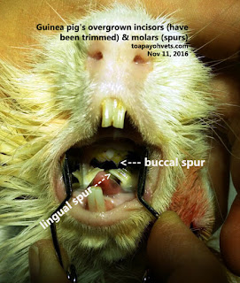
Clipping the front incisors every month for the past 3 months did not help. It is cheap but the lower molars overgrow cutting into the tongue as you can view in the images. The guinea pig eats less. Teeth continuously grow but not worn out by chewing. Less eating. Lose weight. Emaciated. Molar teeth roots mis aligned. Decay. Abscess in lower jaw. Swelling seen in left lower jaw. No stools passed. Will die soon if not treated.
-------------------------------
The below are the updated images in
2020 for the video production
 |
Use the appropriate rodent dental equipment. This rodent cheek dilator is very useful
in opening the mouth wide for a detailed physical examination |
 |
| Gas anaesthesia without injectable anaesthetics is safer for the GP |
--------------------------------
Older images in 2016, not photoshopped well.
.
The mouth cannot be examined properly just by opening it. It needs anaesthesia. But the risk is so high. So, we are back to square one.
NOTES
1. Dental disease. Runny eyes, runny nose, drooling, teeth grinding, selective eating, not eating, weight loss, slobbering.
2. Guinea pig may not have the palpable protrusions at the ventral aspects of the mandible or at the lateral aspects of the maxilla as in the rabbit or chinchilla. Therefore, it may be hard to detect malocclusion.
3. GP + chinchilla
2(I1/1, C0/0, PM 1/1, M3/3) = 20 teeth.
Rabbit has I2/1 in case you don't know. It has 4 obvious front teeth and 2 more shorter and smaller upper front teeth behind the front teeth!


