DRAFT SCRIPT for video production.
HOOK
Use footage 0:00 - 0:55 as hook and narrate the following below.


Tiong Bahru apartments are over 60 years old

Singapore Postman

Tiong Bahru market


 Prognosis for trichoblastoma is good with surgical removal as most do not become cancerous. However, there are rare cases of some being cancerous.


-----------------------------------------------
For the past 11 years, the Silkie Terrier had a small skin lump on her head. It slowly grew bigger over the years.
However, in the past 2 months, it grew much more. It became inflamed. See the big pinkish lump (pause this footage in the video).
The lump had changes in its cell structure as it grew bigger. Hence, it become extremely itchy.
The lump had changes in its cell structure as it grew bigger. Hence, it become extremely itchy.
The owner consulted Dr Daniel of Toa Payoh Vets on Dec 18, 2014.

PAUSE
This is a


Singapore is an island state of around 700 sq km. Small breed dogs are most popular as around 80% of the residents live in high-rise apartments.

Tiong Bahru apartments are over 60 years old
Singapore Postman

Tiong Bahru market
Silkie terriers used to be a favourite breed as pets
in the past 10 years. Skin lumps develop as small papules and then nodules as the pet grows older. 

However, many of the baby boomer generation of owners do not have time to get their pets health checked after puppyhood as in this case study. 

Fruit stall in Tiong Bahru market

"My dog rubs her head tumour against the sofa many times!" the lady owner spoke loudly as was her usual habit. "She just can't stop doing it."
(Animation of dog rubbing sofa?)😢😢😢😢😢
(Animation of dog rubbing sofa?)😢😢😢😢😢
"The head of the lump is inflamed or red now," the vet explained. That inflammation cause itchiness. It can be a benign tumour like a cancerous tumour like basal cell carcinoma or an abscess.

"Surgery to cut it off is the only treatment.
"After cutting out the skin lump, send the lump to the laboratory for histology. Histology is the microscopic examination of the tissue cells to check whether it is cancerous or not.
"After cutting out the skin lump, send the lump to the laboratory for histology. Histology is the microscopic examination of the tissue cells to check whether it is cancerous or not.
"A fast growing tumour in an old dog may be basal cell carcinoma. This is a cancerous tumour."
The lady owner, in her late 40s, consented to the blood test, anaesthesia, surgery and histology.
The lady owner, in her late 40s, consented to the blood test, anaesthesia, surgery and histology.
SURGERY
Use footage 0:00 - 0:32 as you narrate.
https://www.youtube.com/watch?v=dJRFqvtEFW4
https://www.youtube.com/watch?v=dJRFqvtEFW4
The blood test did not show any abnormalities. The dog was fit for anesthesia. Dr Daniel used a scalpel to cut out the skin lump and sent it for histology.

HISTOLOGY is important to check:
1. whether the lump is cancerous or not. It was not cancerous.
2. whether it has had been completely cut out. In this case,
2. whether it has had been completely cut out. In this case,
the surgery completely removed it as Dr Daniel cut with 1-cm margin.
(Show the histology report and read:)
The tumour is completely excised. No overt malignancy is present.
Diagnosis: Suggestive of a trichoblastoma, not involving the resection margin.
Diagnosis: Suggestive of a trichoblastoma, not involving the resection margin.
(Pause the footage to let viewer see report in video).

What is a trichoblastoma?
The trichoblastoma is a benign cutaneous tumor that may derive from the hair germ of a developing follicle. It is common in dogs.
(SHOW AN IMAGE OF IT FROM THE VIDEO FOOTAGE)
(SHOW AN IMAGE OF IT FROM THE VIDEO FOOTAGE)
Trichoblastomas are usually present on the face and head as non-ulcerated nodules or papules. They are very rarely pigmented or cancerous.
Trichoblastoma may be inflamed and become itchy as in this Silkie Terrier.
On histology, trichoblastomas are large circumscribed basaloid tumors, located in the middle to lower dermis, without epidermal connection.
Tips and advices: (narrate image)

Trichoblastic carcinoma may appear similar to trichoblastomas clinically. However, on histology, there is significant asymmetry of the architecture, atypical mitotic figures, necrosis, and substantial infiltration of the tumor into the subcutaneous fat and muscle evident in trichoblastic carcinoma.
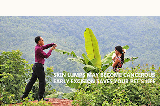
Hence early excision saves your pet's life.
Credits:


-------------------------------------------------------------
Toa Payoh Vets google page images


-------------------------------------------------------------
HISTOLOGY CAN TELL WHETHER THE HEAD NODULE IS CANCEROUS OR NOT.
Basal cell carcinoma and trichoblastic carcinoma may appear similar to trichoblastomas clinically.
Basal cell carcinomas are malignant, proper diagnosis and management is important.
On histology, trichoblastomas are large circumscribed basaloid tumors, located in the middle to lower dermis, without epidermal connection.
The use of immunohistochemistry to show the staining patterns.
Histologically there are retraction spaces, atypia, and single cell necrosis in basal cell carcinoma. In addition, basal cell carcinomas do not stain for cytokeratin 20 and CD34 antigens. They also have diffuse staining of bcl-2 compared to peripheral staining seen in trichoblastomas.
Basal cell carcinomas do not stain for cytokeratin 20 and CD34 antigens. They also have diffuse staining of bcl-2 compared to peripheral staining seen in trichoblastomas.
Basal cell carcinomas do not stain for cytokeratin 20 and CD34 antigens. They also have diffuse staining of bcl-2 compared to peripheral staining seen in trichoblastomas.
-----------------------------------------------
RESEARCH NOTES
TP 45715
Dec 18, 2014 Case consultation and operation videos
Dog, F, 11 yrs
Dec 18, 2014 Case consultation and operation videos
Dog, F, 11 yrs
Pt 1
Difficult to give tablet after surgery - excision of skin lump by Dr Daniel today. It is best to get small skin lump removed by your vet as some of them do turn malignant or irritate the dog. This dog was rubbing the skin tumour above the neck against the bed etc. for many years.
https://www.youtube.com/watch?v=LfEGOUBbHGs
Pt 2
https://www.youtube.com/watch?v=dJRFqvtEFW4
Op
Pt 3. Post op.
https://www.youtube.com/watch?v=Wu8jDyhyg2w
Pt 2
https://www.youtube.com/watch?v=dJRFqvtEFW4
Op
Pt 3. Post op.
https://www.youtube.com/watch?v=Wu8jDyhyg2w
Difficult to give tablet after surgery - excision of skin lump by Dr Daniel today. It is best to get small skin lump removed by your vet as some of them do turn malignant or irritate the dog. This dog was rubbing the skin tumour above the neck against the bed etc. for many years.
No comments:
Post a Comment
Note: Only a member of this blog may post a comment.