https://youtu.be/Sqz2dCNO4iU Edit footage to give a concise account
--------------------------------
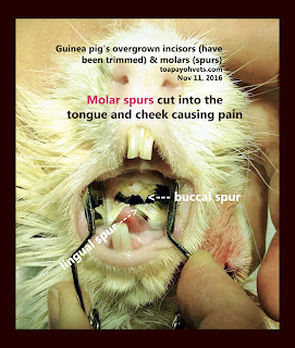
Overgrown front teeth (incisors) had been trimmed. No images were taken, but the video shows
the process.
Extraction of the abscessed molars would be done 7 days later if the guinea pig is fit for anaesthesia.
I had drained and lanced the left jaw abscess. It was filled with foul-smelling pus.
The owner did not return for dental work.
--------------------------------------------
In December 2015, the guinea pig had overgrown incisors and molar spurs clipped short by Dr Sing Kong Yuen one year ago and had good appetite. No dental X-rays were taken as the owner wanted the least cost.
From July - September 2016, the father and son would bring the guinea pig to cut short the overgrown front teeth only every month. This was an inexpensive procedure compared to more detailed dental X-rays. The guinea pig would eat again.
But in Nov 8, 2016, they consulted me as the 5-year-old guinea pig was emaciated, anaemic and was not eating. There was a gigantic left jaw abscess. Two treatment options are available.
1. Marsupialisation - opening up a big hole, drain the abscess and stitch the mucosa (inside layer) of the abscess to the skin, creating an open wound. The wound can be flushed daily.
2. Remove the capsule of the encapsulated abscess. This would permit the pus to be drained
At Toa Payoh Vets, I incised and drain the abscess. The decayed molar tooth can be extracted when the guinea pig is in better health. The owner has to return for this dental work. The owners did not return for follow-up.
In conclusion, three procedures are needed when the guinea pig has overgrown front teeth and is not eating and loses weight.
1. Physical examination for jaw abscess and dental malocclusion
2. Examination of molar spurs using the otoscope.
3. Skull X-rays, using oblique views to see the teeth.


No comments:
Post a Comment
Note: Only a member of this blog may post a comment.