Oct 7, 2018, I have updated the webpage written in 2011. The webpage is:
The above webpage is reproduced below:
 TOA
PAYOH VETS TOA
PAYOH VETStoapayohvets.com Date: 07 October, 2018 Focus: Small animals - dogs, cats, hamsters, guinea pigs, turtles & rabbits
| |||||||||||||||||
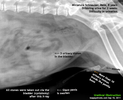

4. Consider hydrochlorothiazide (2mg/kg q 12 hr) with highly recurrent urolithiasis in dogs without hypercalcaemia.
5. Consider potassium citrate (75mg/kg q 12-24 hr) if urine pH is consistently <6 .5.="" br="">
CONCLUSION
Many dog owners in Singapore stop doing regular X-rays and urine tests, leading to recurrence of urinary stones and surgeries or euthanasia. Feeding Hills' C/D is not advised in calcium oxalate urolithiasis as it is an urine acidifying diet. There is no Prescription Diet to dissolve calcium oxalate stones.
In Singapore in 2018, Hills' C/D diet is no longer available. Royal Canin still produces Urinary S/0 diets. These diets are acidifying diets used mainly for struvite stone prevention.
It is best to consult your veterinarian to prevent recurrence of calcium oxalate urolithiasis as each case is different. E-mail judy@toapayohvets.com or SMS +65 9668-6468 to make an appointment for consultation of your case or for a second opinion.
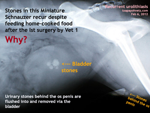
FOLLOW-THROUGH in Feb 2012
Recurrent urolithiasis 4 months later - struvite stone case study. This is not a calcium oxalate stone case study.
Advertisement: Asia USA Realty (Singapore)
asiahomes.com Pte Ltd
VARIOUS UNITS ARE FOR RENT AT VARIOUS TIMES:
Tel: +65 9668 6468, Email: judy@asiahomes.com
Asia USA Realty
asiahomes.com Pte Ltd
VARIOUS UNITS ARE FOR RENT AT VARIOUS TIMES:
Tel: +65 9668 6468, Email: judy@asiahomes.com
Asia USA Realty
 More
info at:
Dogs or
Cats. To make an appointment:
e-mail
judy@toapayohvets.com
More
info at:
Dogs or
Cats. To make an appointment:
e-mail
judy@toapayohvets.comtel: +65 6254-3326 or 9668-6469
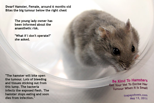 Hamster anaesthesia at Toa
Payoh Vets
Hamster anaesthesia at Toa
Payoh Vets
To
make an appointment: e-mail
judy@toapayohvets.com
tel: +65 9668-6469, 6254-3326
tel: +65 9668-6469, 6254-3326
 toapayohvets.com
Be Kind To Pets
Veterinary Education
Project 2010-0129
|
Toa
Payoh Vets Clinical Research |
Copyright ©
Asiahomes
All rights reserved. Revised: October 07, 2018
Toa Payoh Vets
-------------------------------------------------------------------------------------------------------------------------------------
,30 Extracorporeal shockwave lithotripsy has a low mortality rate (<2 15="" a="" and="" but="" concurrent="" dogs.="" dogs="" extracorporeal="" for="" in="" is="" larger="" lithotripsy="" nephroliths.="" obstruction="" of="" placement="" recommended="" requires="" retreatment="" shockwave="" span="" stent="" style="box-sizing: border-box;" the="" typically="" undergoing="" ureteral="" ureteroliths="" with="">13
All rights reserved. Revised: October 07, 2018
Toa Payoh Vets
-------------------------------------------------------------------------------------------------------------------------------------
University of Minnesota Urolith Centre
1. TOP 10 CANINE CALCIUM OXALATE UROLITH FORMING BREED BY CONTINENT
BASED ON DATA 2013-2017
BASED ON DATA 2013-2017
- Asia
1.Shih Tzu 2.Miniature Schnauzer 3.Maltese 4.Chihuahua 5.Mixed 6.Pomeranian 7.Yorkshire Terrier 8.Papillon 9.Toy Poodle 10.Miniature Poodle
2. Early recognition of CaOx stones include nonsurgical removal, early dietary modification to minimize urolith growth, and early removal to avoid urinary obstruction.
3. Globally, calcium oxalate (CaOx) is the most frequent urinary stone affecting dogs.
4. High-risk dogs
Males are affected more than females. Around the world the most common breeds that form calcium oxalate are the Maltese, miniature Schnauzer, Shih Tzu, Yorkshire terrier, and Chihuahua. The most common age for the onset of clinical disease is 7 to 8 years. We recommend to begin screening (i.e. medical imaging) high risk dogs at 5 to 6 years of age.
5. X-ray advices
Why you see them, why you don't. (Radiographs and Uroliths)
Don’t be fooled by published texts describing urate and cystine uroliths as radiolucent. They are correct that urate and cystine are the least radiopaque of the common stones in dogs and cats. However, radiographic appearance of uroliths depends on several factors of which size and mineral type are the most important. Small urate and cystine stones (Figure 1 and 2) may be radiographically difficult to discern but as they become larger they are more obvious (Figures 2 and 3).
Things that can be done to improve radiographic discernibility
1. A full bladder improves contrast 2. A steady patient improves contrast 3. Imaging the entire urinary tract avoids missing urethral and kidney stones 4. Pulling the legs back or forward away from the proximal os penis prevents femurs from obscuring stones 5. When in doubt additional imaging techniques may be needed (e.g. contrast enhanced radiography, ultrasonography, computerized tomography) 6. Look at extra-urinary structures (dogs with small livers may be an indication of urate stones in dogs with portovascular shunts).
6. Stone analysis. Rings of growth similar to those in a tree
https://www.vetmed.umn.edu/sites/vetmed.umn.edu/files/rings_of_a_stone.pdf
The Rings of Saturn – Minnesota Urolith Center, Image of the Month, Nov 2017 Tree-ring dating, dendrochronology, is the study of climate and environmental changes as recorded by the growth rings of a tree. These concentric rings can be wide, indicating a wet season of rapid growth, narrow indicating a dry season of slow growth, or blemished, indicating fire or insect damage (Figure 1). A similar analogy, urochronology, can be used to tell the life-story of uroliths. Figure 2 is a urolith from Saturn, a female/spayed, Jack Russell terrier. The center or origin of the urolith is calcium oxalate indicating excessive urinary calcium excretion (hypercalciuria) resulting from metabolic, genetic, dietary or idiopathic causes. The white layer is struvite indicating urinary tract infection caused by urease producing bacteria. Staphylococcus and Proteus spp. are common examples. Next, a dark narrow band of calcium oxalate is repeated indicating a period of antibiotic clearance of bacteria but without control of hypercalciuria. Finally, the struvite returns in the outer layer because antibiotics were prematurely stopped. Figure 3 is a bladder stone from a female Labrador retriever. The center is composed of calcium phosphate carbonate and the remaining outer layers are struvite. Both minerals are infection induced. The wide layers likely represent periods of rapid growth and the narrow layers are periods of slower growth. Reading the layers of bladder stones provide a glimpse into the climate in the urinary tract during formation. This story of urochronology can help determine therapy needed to minimize recurrence.
7. Prevention and Treatment
1. Alkalinising therapeutic diet. Monitoring using urine test monthly and regular radiographs (6-12 monthly)
2. Prevent hypercalciuria. Urine test to check whether the urine is acidic. Calcium oxalate uroliths form in acidic urine.
3. Voiding urohydropropulsion is possible if sizes are small (less than 5 mm). Early detection by lateral radiograph/ultrasound every 6-12 monthly.
4. Laser
--------------------------------------------------------------------------------
Case study
Duking out the treatment of Struvite/Urate Urolithiasis "Duke" is a 12-yr-old MN Shih Tzu presented for intermitted bloody urine. Antibiotics initially resolved clinical signs, but hematuria recurred. Radiographs identified a moderately radiopaque bladder stone (Figure 1). "Duke" was anesthetized, the stone was fragmented by laser lithotripsy (video 1), and the fragments were removed by basket retrieval and voiding urohydropropulsion. Quantitative analysis of the stone was 70% struvite and 30% urate (Figure 2). Taking a second look at the radiograph, liver size was normal but small. Serum concentrations of liver enzymes were unremarkable: ALP = 28 U/L (normal = 22-92), AST U/L = 22 (normal = 16-44) and ALP U/L = 25 (normal = 8 to 139). How would you manage this dog?
Diagnostically- 1. Shih Tzu's are predisposed to porto-vascular shunts. Consider evaluating bile acids in breeds at risk for liver shunts. Postprandial serum bile acid concentration was high (52 µmol/L; normal is 15 to 25). 2. Culture urine, struvite uroliths in dogs are usually due to a urinary tract infection.
Therapeutically- 1. To prevent urate, consider canned lower purine/protein foods usually formulated with egg, dairy, or vegetable proteins that result in a more neutral or alkaline urine pH (e.g. Hill's l/d, u/d, i/d Sensitive Canine, HA Hydrolyzed, others). 2. To prevent struvite, control urinary tract infection with periodic urine cultures and appropriate antimicrobics, when needed.
If bile acids were normal, we would still avoid urine acidification (a common feature of struvitolytic diets) because urate is more soluble in alkaline urine and therefore less likely to precipitate out.
--------------------------------------------------------------------
Voiding Urohydropropulsion—Raincoat Required ? Image of the Month, June 2017
At the Minnesota Urolith Center, we developed voiding urohydropropulsion (VU), a nonsurgical method of removing small bladder stones from dogs and cats. Stone removal typically takes a few seconds and is successful, compassionate alternative to surgical stone removal. Voiding urohydropropulsion requires lifting the anesthetized animal into a vertical position, and can be cumbersome in large dogs. To assist with the procedure, we use a tilting exam table. Video of voiding urohydropropulsion in a 6 year old, male-neutered dalmatian using a tilting exam table.
Additional Resources:
Use of urohydropropulsion, cystoscopy and lithotripsy to manage feline urolithiasis, Jody Lulich DVM, PhD, DACVIM, Hill's Global Symposium on Feline Lower Urinary Tract Health, Prague, 23rd - 24th April 2014 Voiding urohydropropulsion. Lessons from 5 years of experience. Lulich, JP, et al, Vet Clin North Am Small Anim Pract. 1999 Jan;29(1):283-91, xiv.
------------------------------------------------
More than just a pretty picture Minnesota Urolith Center Case of the Month December, 2016 This lateral radiograph (figure 1) from an adult Labrador retriever reveals a radiopaque urocystolith with radiating spokes. this was an incidental finding during a workup for gastroenteritis. What is your prediction of the stone's mineral composition?
Its radiographic appearance is consistent with silica, which was confirmed by quantitative analysis. (figure 2). But this radiograph offers more than just a clue to the urolith composition. Silica is the most abundant mineral on the surface of the earth. The owner reports the dog does not eat soil. Take another look at the radiograph and decide for yourself. The radiopaque material in the colon is consistent with rocks or other high density mineral material.
Learn more about the radiographic prediction of stone type with our links below. On our App is a CALCulator function to assist prediction of urolith composition. View our recommendations to learn about dietary and pharmaceutical sources of silica that may contribute to silica stone formation along with a plan to prevent stone recurrence.
https://www.vetmed.umn.edu/centers-programs/minnesota-urolith-center/recommendations
------------------------------
https://onlinelibrary.wiley.com/doi/full/10.1111/jvim.14559
CATS
3. Globally, calcium oxalate (CaOx) is the most frequent urinary stone affecting dogs.
4. High-risk dogs
Males are affected more than females. Around the world the most common breeds that form calcium oxalate are the Maltese, miniature Schnauzer, Shih Tzu, Yorkshire terrier, and Chihuahua. The most common age for the onset of clinical disease is 7 to 8 years. We recommend to begin screening (i.e. medical imaging) high risk dogs at 5 to 6 years of age.
5. X-ray advices
Why you see them, why you don't. (Radiographs and Uroliths)
Don’t be fooled by published texts describing urate and cystine uroliths as radiolucent. They are correct that urate and cystine are the least radiopaque of the common stones in dogs and cats. However, radiographic appearance of uroliths depends on several factors of which size and mineral type are the most important. Small urate and cystine stones (Figure 1 and 2) may be radiographically difficult to discern but as they become larger they are more obvious (Figures 2 and 3).
Things that can be done to improve radiographic discernibility
1. A full bladder improves contrast 2. A steady patient improves contrast 3. Imaging the entire urinary tract avoids missing urethral and kidney stones 4. Pulling the legs back or forward away from the proximal os penis prevents femurs from obscuring stones 5. When in doubt additional imaging techniques may be needed (e.g. contrast enhanced radiography, ultrasonography, computerized tomography) 6. Look at extra-urinary structures (dogs with small livers may be an indication of urate stones in dogs with portovascular shunts).
6. Stone analysis. Rings of growth similar to those in a tree
https://www.vetmed.umn.edu/sites/vetmed.umn.edu/files/rings_of_a_stone.pdf
The Rings of Saturn – Minnesota Urolith Center, Image of the Month, Nov 2017 Tree-ring dating, dendrochronology, is the study of climate and environmental changes as recorded by the growth rings of a tree. These concentric rings can be wide, indicating a wet season of rapid growth, narrow indicating a dry season of slow growth, or blemished, indicating fire or insect damage (Figure 1). A similar analogy, urochronology, can be used to tell the life-story of uroliths. Figure 2 is a urolith from Saturn, a female/spayed, Jack Russell terrier. The center or origin of the urolith is calcium oxalate indicating excessive urinary calcium excretion (hypercalciuria) resulting from metabolic, genetic, dietary or idiopathic causes. The white layer is struvite indicating urinary tract infection caused by urease producing bacteria. Staphylococcus and Proteus spp. are common examples. Next, a dark narrow band of calcium oxalate is repeated indicating a period of antibiotic clearance of bacteria but without control of hypercalciuria. Finally, the struvite returns in the outer layer because antibiotics were prematurely stopped. Figure 3 is a bladder stone from a female Labrador retriever. The center is composed of calcium phosphate carbonate and the remaining outer layers are struvite. Both minerals are infection induced. The wide layers likely represent periods of rapid growth and the narrow layers are periods of slower growth. Reading the layers of bladder stones provide a glimpse into the climate in the urinary tract during formation. This story of urochronology can help determine therapy needed to minimize recurrence.
7. Prevention and Treatment
1. Alkalinising therapeutic diet. Monitoring using urine test monthly and regular radiographs (6-12 monthly)
2. Prevent hypercalciuria. Urine test to check whether the urine is acidic. Calcium oxalate uroliths form in acidic urine.
3. Voiding urohydropropulsion is possible if sizes are small (less than 5 mm). Early detection by lateral radiograph/ultrasound every 6-12 monthly.
4. Laser
--------------------------------------------------------------------------------
Case study
Duking out the treatment of Struvite/Urate Urolithiasis "Duke" is a 12-yr-old MN Shih Tzu presented for intermitted bloody urine. Antibiotics initially resolved clinical signs, but hematuria recurred. Radiographs identified a moderately radiopaque bladder stone (Figure 1). "Duke" was anesthetized, the stone was fragmented by laser lithotripsy (video 1), and the fragments were removed by basket retrieval and voiding urohydropropulsion. Quantitative analysis of the stone was 70% struvite and 30% urate (Figure 2). Taking a second look at the radiograph, liver size was normal but small. Serum concentrations of liver enzymes were unremarkable: ALP = 28 U/L (normal = 22-92), AST U/L = 22 (normal = 16-44) and ALP U/L = 25 (normal = 8 to 139). How would you manage this dog?
Diagnostically- 1. Shih Tzu's are predisposed to porto-vascular shunts. Consider evaluating bile acids in breeds at risk for liver shunts. Postprandial serum bile acid concentration was high (52 µmol/L; normal is 15 to 25). 2. Culture urine, struvite uroliths in dogs are usually due to a urinary tract infection.
Therapeutically- 1. To prevent urate, consider canned lower purine/protein foods usually formulated with egg, dairy, or vegetable proteins that result in a more neutral or alkaline urine pH (e.g. Hill's l/d, u/d, i/d Sensitive Canine, HA Hydrolyzed, others). 2. To prevent struvite, control urinary tract infection with periodic urine cultures and appropriate antimicrobics, when needed.
If bile acids were normal, we would still avoid urine acidification (a common feature of struvitolytic diets) because urate is more soluble in alkaline urine and therefore less likely to precipitate out.
--------------------------------------------------------------------
Voiding Urohydropropulsion—Raincoat Required ? Image of the Month, June 2017
At the Minnesota Urolith Center, we developed voiding urohydropropulsion (VU), a nonsurgical method of removing small bladder stones from dogs and cats. Stone removal typically takes a few seconds and is successful, compassionate alternative to surgical stone removal. Voiding urohydropropulsion requires lifting the anesthetized animal into a vertical position, and can be cumbersome in large dogs. To assist with the procedure, we use a tilting exam table. Video of voiding urohydropropulsion in a 6 year old, male-neutered dalmatian using a tilting exam table.
Additional Resources:
Use of urohydropropulsion, cystoscopy and lithotripsy to manage feline urolithiasis, Jody Lulich DVM, PhD, DACVIM, Hill's Global Symposium on Feline Lower Urinary Tract Health, Prague, 23rd - 24th April 2014 Voiding urohydropropulsion. Lessons from 5 years of experience. Lulich, JP, et al, Vet Clin North Am Small Anim Pract. 1999 Jan;29(1):283-91, xiv.
------------------------------------------------
More than just a pretty picture Minnesota Urolith Center Case of the Month December, 2016 This lateral radiograph (figure 1) from an adult Labrador retriever reveals a radiopaque urocystolith with radiating spokes. this was an incidental finding during a workup for gastroenteritis. What is your prediction of the stone's mineral composition?
Its radiographic appearance is consistent with silica, which was confirmed by quantitative analysis. (figure 2). But this radiograph offers more than just a clue to the urolith composition. Silica is the most abundant mineral on the surface of the earth. The owner reports the dog does not eat soil. Take another look at the radiograph and decide for yourself. The radiopaque material in the colon is consistent with rocks or other high density mineral material.
Learn more about the radiographic prediction of stone type with our links below. On our App is a CALCulator function to assist prediction of urolith composition. View our recommendations to learn about dietary and pharmaceutical sources of silica that may contribute to silica stone formation along with a plan to prevent stone recurrence.
https://www.vetmed.umn.edu/centers-programs/minnesota-urolith-center/recommendations
------------------------------
https://onlinelibrary.wiley.com/doi/full/10.1111/jvim.14559
CATS
Recommendation 2.3: Dissolution Should not be Attempted in Cats with Obstructive Upper Urinary Tract Uroliths
Rationale: Over 90% of nephroliths and ureteroliths in cats are composed primarily of calcium oxalate. Calcium oxalate uroliths are not amenable to medical dissolution. Delaying appropriate care may contribute to an irreversible decrease in kidney function.31-33
------------------
Medical Treatment for Obstructive Ureterolithiasis is Rarely Effective, Consider Minimally Invasive Removal
Medical management of stable obstructive ureterolithiasis can be considered for 24–72 hours. However, clients should be informed of the high rate of medical failure. Medical treatment should include fluid diuresis and mannitol continuous rate infusion treatment, if tolerated. Alpha adrenergic antagonists and tricyclic antidepressants also have been used with anecdotal reports of improvement in some cases and can be considered if not contraindicated. Medical treatment should not be continued in animals that are persistently oliguric or anuric, hyperkalemic, have progressive azotemia and progressive renal pelvic dilatation; minimally invasive urolith extraction or bypass is needed. Fluid treatment should be closely monitored to prevent overhydration. In dogs, in addition to propulsive treatment for uroliths, broad‐spectrum antimicrobials IV (ideally for at least 24 hours before intervention) should be administered.
Ureterolith‐induced ureteral obstructions should be monitored rather than decompressed when renal pelvic dilatation is ≤3–5 mm, and renal function is stable. Intervention only should be considered when an experienced operator is available.
Rationale: Medical management for the treatment of cats with ureteral obstructions is only reported to be effective in 8–13% of cases.31 Because over 25% of ureteral obstructions in cats are associated with concurrent ureteral strictures, success of medical management often is limited.33 In dogs, 59% of all ureteral obstructions, and 85% of those with pyonephrosis, had evidence of UTI at the time of diagnosis, supporting the administration of antimicrobials.2,30
Higher complication rates are seen with less experienced operators. This may affect timing of surgical intervention, and waiting for a more experienced operator is ideal for the best possible outcome.
Recommendation 2.8: Obstructive Ureteroliths in Cats should be Managed by Subcutaneous Ureteral Bypass or Ureteral Stenting
Subcutaneous ureteral bypass or ureteral stenting for ureteral obstructions in cats should be considered the first choice for the best possible outcome. We emphasize that fluoroscopic imaging, proper training, and an experienced operator are needed to optimize the patient outcomes.
Recommendation 2.9: Obstructive Ureteroliths in Dogs should be Managed by Ureteral Stenting
Ureteral stents are the treatment of choice for ureterolith‐induced ureteral obstructions in dogs if performed by a trained operator. This approach may be combined with subsequent extracorporeal shockwave lithotripsy if necessary.13 Interventional options such as ureteral stent placement, extracorporeal shockwave lithotripsy, or both for the treatment of ureteral obstructions in dogs always should be considered and offered to clients.
Rationale: Ureteral stents are associated with the lowest short‐ and long‐term morbidity and mortality rates when compared to all other reported treatment options.2,30
The morbidity and mortality rates (<2 a="" after="" also="" and="" are="" azotemia="" been="" class="noteLink scrollableLink" dogs="" for="" have="" href="https://onlinelibrary.wiley.com/doi/full/10.1111/jvim.14559#jvim14559-note-1003" improvement="" in="" lower="" of="" placement.="" rate="" reobstruction="" reported="" severity="" stent="" stenting="" style="background-color: transparent; box-sizing: border-box; color: #2f7bae; display: inline; font-size: 13.93px; font-style: normal; margin-left: -3px; padding-bottom: 5px; padding-left: 0px; padding-right: 0px; padding-top: 5px; padding: 5px 0px; position: relative; text-decoration: none; top: -7px;" surgery.="" than="" the="" those="" title="Link to note" traditional="" ureteral="">2
Recommendation 2.10: Ureterolith Composition will Affect Management Decisions
Careful assessment of urinalysis (eg, crystals, urine pH), urine culture results, radiographic appearance, and when possible, quantitative urolith analysis should always be performed. In dogs, suspected struvite ureteroliths should be stented and then dissolved as discussed in the lower urinary tract urolith section. Suspected obstructed calcium oxalate ureteroliths should be either stented for long‐term treatment or stented with concurrent or subsequent extracorporeal shockwave lithotripsy, if necessary. Cystine and urate ureteroliths should be treated by a ureteral stent and concurrent medical and dietary treatment.
Rationale: Ureteral stents in dogs often can be placed endoscopically. This procedure can relieve an emergency situation both effectively and safely. Forty‐four dogs that underwent ureteral stent placement had their stents in place for up to 1158 days, suggesting that long‐term stent placement is possible.2 Owners should be aware of the primarily reobstruction risks that are most often associated with concurrent ureteral stricture. If necessary, a ureteral stent exchange can be performed on an outpatient basis, but is not required for most dogs. Knowing the urolith composition will help by employing appropriate medical and dietary treatment to prevent stent encrustation and future urolith formation. If stenting fails, other options such as extracorporeal shock wave lithotripsy and subcutaneous ureteral bypass device placement, or traditional surgery, can be considered.
Recommendation 2.11: Routinely Culture Urine of Dogs with Ureteral Obstruction and Consider Antimicrobial treatment
Dogs with ureteral obstruction should have their urine cultured and should be given antimicrobial treatment at the time of diagnosis because of the high incidence of concurrent UTI and pyonephrosis.
Rationale: Fifty‐nine percent of 44 dogs with ureteral obstructions had positive urine culture results at the time of diagnosis; 30% had a diagnosis of pyonephrosis and associated sepsis.
------------------------------------------------------
Part 3
Urolith Prevention
Removal or bypass of uroliths will not alter the underlying conditions responsible for their formation. Therefore, it is logical to assume that additional therapeutic strategies are needed to prevent urolith recurrence. The most effective prevention strategies are those that eliminate the underlying cause. For cases in which a cause remains elusive or cannot be altered, minimizing pathophysiologic risk factors associated with formation should be considered.
Nutritional treatment remains a subject of much clinical interest and debate in the management of urolithiasis because of epidemiological and pathophysiological data associating nutrient intake with urine saturation and potential lithogenicity. For some urolith types, nutritional prevention plays a primary role (eg, sterile struvite uroliths), and for other urolith types, nutritional treatment plays a minor role (eg, infection‐induced struvite and infection‐induced calcium phosphate carbonate uroliths). For all mineral types (except infection‐induced struvite), feeding diets high in moisture is one of the cornerstones of urolith prevention strategies.
Recommendation 3.1: Prevent Sterile Struvite Uroliths by Feeding Therapeutic Maintenance Foods with Low Magnesium and Phosphorus that Acidify Urine
Successful prevention of struvite uroliths is dependent on classifying them as sterile or infection‐induced. To make this distinction, aerobic bacterial urine or urolith culture should be performed before antimicrobial treatment. Sterile struvite uroliths, which most often occur in cats, are best prevented by feeding therapeutic maintenance foods with low magnesium and phosphorus that acidify the urine.
Recommendation 3.2: Primary Prevention of Infection‐Induced Struvite Uroliths is Persistent Elimination of Urinary Tract Infection
Primary treatment for preventing infection‐induced struvite uroliths, which is the most common struvite urolith in dogs, is early identification and elimination of UTI. Urine sediment evaluation and pH monitoring are not suitable diagnostic substitutes for aerobic bacterial urine cultures. Second tier treatment to manage infection‐induced struvite uroliths includes therapeutic maintenance foods with low magnesium and phosphorus that acidify the urine.
Rationale: Urinary tract infection with urease‐producing microorganisms is essential for the formation of infection‐induced struvite uroliths. Eliminating these infections will prevent recurrence of infection‐induced struvite uroliths. Therefore, structural and functional risk factors for UTI should be diagnosed and eliminated, and recurrent infection should be monitored in urine. Routine urinalysis is an insensitive marker for UTI.52, 53 Urine should be cultured monthly for 2–3 months and then as clinically indicated based on clinical signs and patient risk factors. Foods marketed to treat struvite urolithiasis will not prevent their recurrence but may delay or minimize, urolith burden in the presence of unrecognized UTI.
Recommendation 3.3: To Minimize Calcium Oxalate Urolith Recurrence, Decrease Urine Concentration, Avoid Urine Acidification, and Avoid Diets with Excessive Protein Content
Calcium oxalate urolithiasis in dogs and cats appears to be driven primarily by hypercalciuria in association with either hypercalcemia (eg, primary hyperparathyroidism, idiopathic hypercalcemia in cats) or normocalcemia.54 Intrinsic risk factors should be evaluated in all patients that have been diagnosed with calcium oxalate uroliths (eg, evaluate serum ionized and total calcium concentrations, parathyroid hormone), and further diagnostic testing should be pursued if clinically indicated.
Selection of effective preventative treatment is challenging because (1) properly designed clinical trials evaluating urolith recurrence have not been published, (2) the exact mechanisms underlying calcium oxalate urolith formation are not completely understood, (3) associative factors identified in epidemiological studies have not been proven to result in disease, and (4) surrogate endpoints of therapeutic efficacy such as relative supersaturation are mathematical models that may not correlate well with calcium oxalate urolith formation.
Rationale: The high recurrence rate of calcium oxalate uroliths warrants a comprehensive approach and regular monitoring. High‐moisture (>75% water) foods should be recommended. Alternatively, sufficient water can be added to dry kibble to increase moisture intake. Strive to achieve a urine specific gravity ≤1.020 in dogs and <1 .030="" 1.1="" 12="" 13="" 2.3="" 73="" 8="" achieve="" additional="" and="" calcium="" cats="" clinically="" concentrations="" consumption="" containing="" days="" decreased="" dogs="" effective="" foods="" for="" from="" high="" ie="" in="" indicated="" lower="" moisture="" more="" normal="" of="" oxalate.="" oxalate="" prevention.="" provides="" quantities="" relative="" short="" significantly="" span="" studies="" style="box-sizing: border-box;" supersaturation="" term="" that="" the="" theoretically="" to="" urine="" water="">55-57
Diets and medications designed to promote urine acidification (pH < 6.5) should be avoided. Diets that promote the formation of acidic urine in dogs (pH < 6.6) and cats (pH < 6.25) were associated with calcium oxalate urolith formation.58-61 In a study of normal cats, calcium oxalate relative supersaturation linearly decreased with increasing urinary pH.51
Ingestion of foods that contain high quantities of animal protein (>10 g/100 kcal) contributes to calcium oxalate uroliths by increasing urine calcium excretion and decreasing urine citrate excretion. Increasing dietary protein from 35% to 57% (dry matter) increased urine calcium concentration by 35% and decreased urine citrate concentration by 45% in cats.62
In dogs and cats with hypercalcemia, correcting or controlling hypercalcemia aids in preventing calcium oxalate urolith recurrence. Doing so is difficult in cats with idiopathic hypercalcemia and no single treatment has been shown to be effective, including glucocorticoids, bisphosphonate administration, or dietary modification using a high‐fiber diet with potassium citrate administration, but 5 cats with idiopathic hypercalcemia had normalization of blood calcium concentrations when treated with a high‐fiber diet.63
Recommendation 3.3a: Feeding High‐Sodium (>375 mg/100 kcal) Dry Foods should not be a Recommended as a Substitute for High‐Moisture Foods
Rationale: High‐sodium foods increase urinary water excretion, but the effects appear to be short‐lived (ie, 3–6 months).64-66 Although the extent of water intake and urine dilution achieved with increased dietary salt might not be similar to that observed with high‐moisture foods, it can be considered in dogs and cats in which owners decline to feed high‐moisture foods.
Recommendation 3.3b: Consider Potassium Citrate or Other Alkalinizing Citrate Salts for Dogs and Cats with Persistently Acidic Urine
Potassium citrate is an alkalinizing salt that when administered PO and metabolized promotes the excretion of more beneficial alkaline urine. Alkaline urine also enhances urinary citrate excretion, and citrate is a chelator of calcium ions.
Rationale: Oral administration of granular potassium citrate (150 mg/kg/d) was associated with variable increased urinary citrate concentration (3 ± 9 mmol/L) compared to a noncitrate control (0.1 ± 0.06 mmol/L).67 This result may have occurred because the optimal dose of citrate has yet to be determined. In a summary of 5 studies with 283 human calcium oxalate stone formers, 97 (34%) reformed stones or had residual stones grow; this outcome occurred in 15% of patients receiving citrate salts compared to 52% of those not receiving citrate.68 One in vitro study demonstrated that citrate, at a concentration similar to that achieved in urine (5 mmol/L), significantly dissolved and detached calcium oxalate monohydrate crystals from Madin‐Darby canine kidney cells.69
Recommendation 3.3c: Consider Thiazide Diuretics for Frequently Recurrent Calcium Oxalate Uroliths
Thiazide diuretics enhance the renal tubular reabsorption of filtered calcium. They also may indirectly affect intestinal calcium absorption and bone calcium deposition. Some recommend the concomitant administration of potassium citrate because thiazide diuretics contribute to urine acidification. We recommend monitoring urine pH first to assess whether or not potassium citrate is needed.
Rationale: A 55% decrease in urinary calcium concentration was reported in urolith‐forming dogs that were treated with hydrochlorothiazide at a dosage of 2 mg/kg q12h.70 A 65% decrease in urinary calcium oxalate relative supersaturation was reported in clinically normal cats receiving hydrochlorothiazide at a dosage of 1 mg/kg q12h.71
Recommendation 3.4: to Minimize Urate Urolith Recurrence, Decrease Urine Concentration, Promote Alkaline Urine, and Limit Purine Intake
Hyperuricosuria, concentrated urine, and acidic urine are the predominant factors driving urate urolith formation. In most dogs and cats, uric acid, an intermediate product of purine metabolism, is transported to the liver where it is further metabolized by intracellular hepatic uricase to allantoin, an innocuous nitrogenous compound with relatively high water solubility. Defective uric acid transporters (ie, SLC2A9 genetic mutation) and hepatic porto‐vascular anomalies have been identified as common causes for hyperuricosuria and subsequent urate urolithiasis. However, in some animals, particularly cats, the causes for hyperuricosuria and urate urolith formation remain idiopathic.
For dogs with the SLC2A9 mutation (eg, Dalmatians, Bulldogs), urate urolith recurrence can be minimized by increasing fluid intake, promoting alkaline urine (pH ≥ 7), and limiting purine intake. In cats and dogs with porto‐vascular anomalies (eg, Yorkshire terrier, Pug), correcting of the vascular anomaly should also be considered, if appropriate. Data in cats are limited, but purine restriction and urine alkalization are recommended and found to be effective.
Rationale: The high recurrence rate of urate uroliths warrants a comprehensive approach.72 High‐moisture (>75% moisture) foods should be recommended. Alternatively, sufficient water can be added to dry kibble to increase the moisture intake. Strive to achieve a urine specific gravity ≤1.020 in dogs and <1 .030="" achieve="" acid="" additional="" cats.="" concentrations="" consumption="" effective="" in="" lower="" more="" of="" p="" prevention.="" provides="" to="" uric="" urine="" water="">
Urate solubility increases with increasing urine pH. Although the solubility of ammonium urate is thought to plateau at pH ≥ 7.2, in vitro dissolution occurred a high rate at pH ≥ 8.0.73
Dietary purines are precursors of urate and found in virtually all foods. High‐purine foods often are synonymous with high‐protein foods, especially those containing organ meats and fish. Therefore, foods to prevent urate uroliths often are lower in protein. Decreasing dietary protein has been shown to decrease urinary saturation with ammonium urate in healthy dogs.74 Higher‐protein and lower‐purine foods for dogs have also recently been marketed. Selecting an effective food may be difficult because properly controlled studies evaluating urolith recurrence are rare. In 1 study utilizing a crossover design, 6 client‐owned, urolith‐forming Dalmatians were evaluated monthly for urolith recurrence by double‐contrast cystography.4 After 6 months, 50% of dogs consuming the low‐purine and low‐protein prevention food1 developed recurrent uroliths, whereas 87% developed recurrent uroliths while eating the maintenance diet (all recurrent stones were <2 6="" a="" and="" class="noteLink scrollableLink" clinical="" dalmatians="" diameter="" did="" diet="" dogs="" dry="" fed="" forming="" have="" higher="" href="https://onlinelibrary.wiley.com/doi/full/10.1111/jvim.14559#jvim14559-note-1006" in="" low="" mm="" not="" preliminary="" protein="" purine="" results="" signs="" style="background-color: transparent; box-sizing: border-box; color: #2f7bae; display: inline; font-size: 13.93px; font-style: normal; margin-left: -3px; padding-bottom: 5px; padding-left: 0px; padding-right: 0px; padding-top: 5px; padding: 5px 0px; position: relative; text-decoration: none; top: -7px;" title="Link to note" urate="" urolith="">5
Recommendation 3.4.a: Consider Xanthine Oxidase Inhibitors for Dogs Homozygous for Genetic Hyperuricosuria that have Failed Therapeutic Diet Prevention
Rationale: Urate urolith recurrence is common, especially in dogs with a genetic mutation in the urate transporter. Prevention may require more than dietary adjustments. The dosage of allopurinol to sufficiently prevent urate urolith recurrence without xanthine urolith formation is variable and influenced by the severity of disease, endogenous purine production, quantity of purines in the diet, urine pH, and urine volume. In a case series of 10 dogs with previous urate urolithiasis, allopurinol administration in excess of 9–38 mg/kg/d was associated with xanthine urolith formation.76 This occurs because allopurinol inhibits the metabolism of xanthine to uric acid and because xanthine is less soluble in urine than is uric acid. Based on these observations, we recommend a dosage of 5–7 mg/kg q12–24 h to safely prevent urate uroliths.14 The role and effectiveness of allopurinol and newer‐generation xanthine oxidase inhibitors in patients with porto‐vascular shunts are unknown.77 Administration of xanthine oxidase inhibitors should be avoided in dogs that are not receiving decreased purine diets to minimize the risk of xanthine urolith formation. Xanthine oxidase inhibitors have not been formally investigated in cats.
Recommendation 3.5: To Minimize Cystine Urolith Recurrence, Decrease Urine Concentration, Limit Animal Protein Intake, Limit Sodium Intake, Increase Urine PH, and Neuter
Cystinuria is a rare genetic disease that is characterized pathophysiologically by the failure of renal tubular reabsorption of cystine (a poorly soluble amino acid) and phenotypically by highly recurrent cystine urolith formation. Newer classification systems for cystinuria have been published recently.21 Few controlled studies have evaluated the prevention strategies. Lack of clinical information necessitates that therapeutic regimens be monitored frequently and individually adjusted to improve therapeutic efficacy and avoid adverse events.
Rationale: The relative insolubility of cystine in urine and the high recurrence rate warrant a comprehensive approach for urolith prevention. High‐moisture (>75% moisture) foods should be recommended. Alternatively, sufficient water can be added to dry kibble to increase moisture intake. Strive to achieve a urine specific gravity ≤1.020 in dogs and <1 .030="" achieve="" additional="" and="" cats="" concentrations="" consumption="" cystine="" gravities="" improves="" in="" lower="" of="" p="" potentially="" prevention.="" specific="" subsequently="" to="" urine="" water="">
Cystine solubility increases with increasing urine pH.18 In vitro studies that achieved a urine pH > 7.5 increased the efficacy of thiol drugs to solubilize cystine in the urine of cystinuric humans.19 Therefore, potassium citrate or other alkalinizing citrate salts should be administered to dogs and cats with persistently acidic urine. The dosage should be gradually increased to achieve a urine pH of approximately 7.5.
Dietary methionine is a sulfur‐containing amino acid that is precursor of cystine, another sulfur‐containing amino acid. Methionine is common in many animal‐derived nutrients and some plant‐derived nutrients (eg, nuts, tofu, wheat). Diets for the prevention of cystine uroliths should be low in methionine and cystine precursors with adequate amounts of taurine and carnitine. Selecting an effective commercially prepared food may be difficult because controlled studies evaluating stone recurrence have not been reported. Feeding high‐protein diets, particularly those rich in methionine, a cystine precursor, should be avoided in cystinuric dogs. However, the degree of protein restriction that is needed is controversial because protein quality and quantity may affect carnitine content. Carnitine deficiency and associated dilated cardiomyopathy were reported in 5 cystinuric dogs fed low‐protein diets.78 In 1 study performed on cystinuric dogs, the consumption of a decreased protein, urine‐alkalinizing, canned food1 resulted in a 20–25% decrease in 24‐hour urine cystine excretion compared with a canned maintenance diet.17 Canned foods of primarily plant origin also may be helpful in the management of cystine uroliths, but studies documenting the effects on urinary cysteine excretion, urolith prevention, or urolith dissolution are lacking.
In some forms of cystinuria, neutering has been associated with decreases in cystine concentration because of a suspected androgen‐dependent effect, but this effect is not universal.21 Nonetheless, neutering also would prevent unintentional genetic transmission of disease. The effect of neutering on urine cystine concentration in cats has not been investigated.
Recommendation 3.5.A: In Recurrent Cystine Urolith Formers, Add 2‐Mercaptopropionylglycine (Tiopronin, Thiola) to Previously Recommended Prevention Strategies to Further Lower Cystine Concentration and Increase Cystine Solubility
Rationale: Thiol‐binding drugs have been associated with adverse events (eg, fever, anemia, lymphadenopathy). Therefore, they are reserved for patients with more severe disease (nephrolithiasis) or for those with recurrent disease that is not adequately controlled by suitable nutritional and neutering strategies. Thiol‐binding medications work by reducing cystine to 2 cysteine molecules. The thiol‐cysteine product is 50 times more soluble than cystine. Urine alkalization potentiates the effect of thiol‐binding medications.19 Tiopronin is reported to have fewer adverse effects than d‐penicillamine. Tiopronin dosages associated with prevention are 15 mg/kg PO q12h. Because 2‐mercaptopropionylglycine sources are limited, compounding pharmacies have provided this medication for dogs. New compounds disrupting cystine crystal growth (L‐cystine methyl esters) have been proposed.77 Studies in dogs and cats with cystinuria are needed to insure that the efficacy and safety (eg, Fanconi syndrome, kidney failure) profile of these esters is better than that of the current thiol‐binding drugs before their recommended use.
Acknowledgments
Conflict of Interest Declaration: Jody Lulich received competitive research grants related to the consensus statement topic from Hills Pet Nutrition. He participated in multicenter studies evaluating the role of nutrition in the dissolution and prevention of struvite and calcium oxalate uroliths in cats.
Allyson Berent served as paid consultant for Infiniti Medical, LLC, and Norfolk Vet/Access Technology related to the consensus statement topic.
Joseph Bartges serves as associate editor for the Journal of Veterinary Internal Medicine. He was not involved in review of this manuscript.
Larry Adams served as paid consultant related to the consensus statement topic for Hill's Pet Nutrition and sponsorship for continuing education lectures, sponsorship by Royal Canin for continuing education lectures, and sponsorship by Infiniti Medical for continuing education lectures. He conducted paid research for Akina 2010 through 2013, a research project for the development of new ureteral stents.
Carl Osborne in 2016 served as a book section editor of urinary system chapters for “The 5 Minute Veterinary Consult: Canine and Feline” by Larry P. Tilley and Francis W. K. Smith, Jr. He participated in multicenter studies evaluating the role of nutrition in the dissolution and prevention of struvite and calcium oxalate uroliths in cats.
Off‐label Antimicrobial Declaration: The authors declare no off‐label use of antimicrobials.
Footnotes
- 1 Prescription Diet Canine u/d; Hills’ Pet Products, Topeka, KS
- 2 Pavia P, Berent A, Weisse C, et al. Canine Ureteral Stenting for benign ureteral obstruction in dogs. Abstract ACVS, 2014, San Diego, CA
- 3 Berent A, Weisse C, Bagley D et al. Endoscopic nephrolithotomy for treatment of complicated nephrolithiasis in dogs and cats [abstract] ACVS, San Antonio TX, 2013
- 4 Lulich J (abstract), Osborne C, Bartges J, Allen T, et al.: Effects of diets on urate urolith recurrence in Dalmatians. Journal of Veterinary Internal Medicine 1997; 11: 129
- 5 Royal Canin Veterinary Diet Urinary U/C Low Purine Dry Dog Food

No comments:
Post a Comment
Note: Only a member of this blog may post a comment.