Sunday, April 1, 2012

927. E-mail from Kuala Lumpur hamster owner

Claire ...@gmail.com

April 1, 2012 12:23 AM (7 hours ago)


Hi Dr Sing,

I do not know if you will get this mail 'in time' or that I may be too late already.
Either way I feel that I would try anything right now to save my beloved hamster.
I am in Kuala Lumpur, Malaysia.

Ham is 25 months old, very ripe old age.
Started in Nov last year (he was about 18-19 months old), his health started to decline, I presumed due to old age. He suffered some fur loss, his skin had some small black spots and definitely a decline in activities. I did bring him to our local vet here, who did a skin scraping on the black spots to determine if mites or not, and nothing came about. Ham was then diagnosed with a mild Cushing disease, but was advise to just leave him as he is, since he had no problems living his life.

Fast forward to couple of weeks ago, I noticed Ham had some sores/wound on his belly. Large gaping sores, I brought him to a vet again, and was given an antiseptic cleaning lotion and antibiotics. I faithfully cleaned his wounds twice a day and fed him the antibiotics once a day as instructed by the vet. I noticed the sores healed, but not long after, another one popped up.

Early this week, his health took a rapid decline. I thought things were better since the antibiotics was working. But then I noticed he started to sleep daily, did not eat much and lost his eyesight. He became very weak, and frail, and when picked up, he weight very light and was very limp. He does not eat much and I feed him baby food as it is easy to swallow. He still pees, but have not seen him poo. I thought it was his time to come, as he is an old hamster.
What I did not know, I found out today. And I don;t know if I am too late already. I found he had a lump at his neck (throat area)

The thing was when he started to grow older, he developed loose skins around his neck. It was nothing alarming as it was expected when the hamster gets older, but it might have been these loose skin 'masking' the lump until it is affecting him.
The lump us not very large but visible.

My Q is - is it possible for operations? I am asking because I have been googling and found this site: http://www.sinpets.com/F6/20110323massive-neck-tumour-syrian-hamster-surgery-toapayohvets.htm


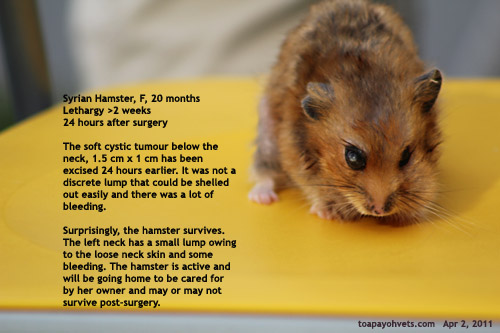
But I am in KL Malaysia, my Ham is weak and not as alert as this ham in the picture.
Please can you advise if surgery is even something to attempt in Ham especially in his condition right now. So far I have yet to properly find any vet here reliable and as caring as I have read in that article above..

If you have advise please help, I am willing to take opportunity if there is, to save him. Many thanks!

E-MAIL REPLY FROM DR SING

I am Dr Sing from Toa Payoh Vets. Thank you for your e-mail. It is very difficult to diagnose by e-mail as NOT all hamster lumps are the same.

In reply, from your description, your hamster is lethargic, not eating and has several skin infected areas (most likely owing to a poor immune system). He unfit for anaesthesia and is very likely to die on the operating table. Definitely he is much older than the Syrian hamster with the neck tumour operated by me.

In conclusion, hamster owners do need to check their hamster for skin lumps daily, even under loose skin or hairless skin. Weigh the hamster weekly to ensure no weight loss. Tumours that are small are easily excised.

However, you may need to find a vet who does operations on hamsters as some vets do NOT perform surgeries, but simply prescribe some medications in the hope that the "lumps" will disappear. Some hamster owners consult the pet shop operators who will sell a "cream" to "shrink" the tumours. Both practices usually do not work unless the lump is a small skin abscess.

Best wishes.


MOST OLDER HAMSTER TUMOURS CAN BE EASILY EXCISED IF THEY ARE SMALL BUT MOST HAMSTER OWNERS WAIT AND SEE OR USE PET SHOP MEDICATION.

Saturday, March 31, 2012

926. Follow-up - The guinea pig that squirted white cloudy urine

http://www.sinpets.com/F6/20120329guinea-pig-websites-singapore_ToaPayohVets.htm
is the website as the following copied report from the above-mentioned website below cannot show the images.


Friday, March 16, 2012
909. Two guinea pigs not eating - white viscous urine, blood in the urine
Yesterday Mar 16, 2012 (Friday), I saw 2 cases of guinea pigs with urinary problems and reviewed the case with Dr Daniel as part of my mentorship program.

We had met an interesting and friendly experienced Australian banker who had travelled to all over the world to work, at Liang Seah Street at 11 am today. "How old are you?" he asked Daniel. He then told Dr Daniel that 20% of the young men of his age and new graduates are jobless in Ireland. He told me that my handshake was weak, compared to Dr Daniel. "Have you been to Timbuktu?" I asked him. "No," he said. "But I have been to various places in Africa. I was sad to see so many homeless children and the poverty."

P.S
I used to study English phases in primary school in the 1960s and there is a phase referring a distant place as Timbuktu which is actually present in Africa. I doubt I have a chance to visit it. One of the guinea pig cases is written and followed up below:

Case of the Guinea Pig squirting out white cloudy urine

March 16, 2012
Not eating. Abdominal swelling obvious. The owner had seen me some months ago. "No more skin infection," she said. "My guinea pig is not eating."
Dr Daniel palpated the swollen abdomen. I did that after him. The swelling covered almost 90% of the abdomen. Soft distended bladder, in my opinion. I don't know what Dr Daniel thought. Was he thinking of ascites? Definitely, ascites present this pendulous abdomen.

Before I could speak further, the guinea pig squirted out white cloudy urine onto the stainless steel consultation table top. "The urine is not normal," I said. "I would take some for urine test." I took a new syringe to suck up some urine. The guinea pig voided much more urine. Thick cloudy liquid. As if the bladder had lots of white sand.

"Is it possible that my guinea pig has bladder stones?" the lady asked. "My other guinea pig had bladder stones. The vet removed it but then said another surgery had to be done."

"Why was there a need for a second surgery?" I asked.
"The first one was not done well," she said. "So, another one had to be done."
"What happened to the guinea pig after bladder surgery?" I asked.
"She lived for a few days and then died."
"In this case, the guinea pig might have or might not have bladder stones. We will wait for a few days and get the urinary tract infection treated first."

A follow up the next 2 - 7 days would be needed. X-rays would be needed to confirm bladder stones.
March 19, 2012
This Guinea Pig was not eating and had a grossly swollen bladder. Dr Daniel and I palpated the abdomen. Looked so much like ascites although I could feel the faint outlines of a very full bladder as big as an orange. Suddenly whitish cloudy urine dribbled out. More and more urine was released like a storm flood. I got the urine inside a syringe and sent to the lab.
Lab Results: Amorphous phosphate crystals +++, bacteria + and urine pH = 9.0.

Evidence of urinary tract infection caused by amorphous phosphate +++. So what to do now?
"In dogs, amorphous phosphate is also known as struvites. Acidify the urine or give S/D diet," I said to Dr Daniel and Dr Vanessa. "What to do for a GP with struvites in the bladder?"

The lady owner returned my text message this morning. She had left all medication to the sister who stays at home. After 3 days, the sister stopped the antibiotics as the GP was eating and drinking.

"But he sits in a corner," the young busy career lady said. "When can I send him down for another check up?" I scheduled next week. She has only one GP and believed that her sister had increased the intake of Vit C tablets to 1.5 tab/day causing the GP to stop eating.

Overdosage of Vit C can lead to kidney stones in people. In this case, the GP had a UTI. Medication was not given for a sufficient number of days.
March 26, 2012
Now, the guinea pig is crated in the Toa Payoh Vets for urine collection (see images below). The urine has been collected and tested again to check for bacterial infection. Bacteria ++ were present. No more amorphous phosphate crystals. I advised oral antibiotics again for 3 days first. The guinea pig was eating normally now.


5153 - 5159. It is good practice to follow up on guinea pigs with urinary tract infections (UTI) as bladder stones could be the cause. Also some owners don't comply with the 7-day instruction to give antibiotics.

Weigh the guinea pig during veterinary consultation. In Singapore, in 2012, the plain-looking guinea pigs are no longer so popular with pet owners!

Friday, March 30, 2012

Bladder stones in a guinea pig - follow-up


Early treatment may or may not save its life. It is hard to guarantee. At this stage, it was unfit for anaesthesia and surgery. I asked the owner to nurse it to good health first. It died the next day, at home.

924. Follow-up: The Guinea Pig that passed fresh blood in the urine

Yesterday, Mar 29, 2012, the lady brought her guinea pig to Dr Daniel at 1pm for consultation as he had passed blood. This guinea pig had been to other vets and we at Toa Payoh Vets was seeing him for the 3rd time today.

First time, I had collected urine for analysis and X-rays.
See case at:
http://www.sinpets.com/F6/20120238guinea-pig-haematuria-singapore_ToaPayohVets.htm


"But no blood in the urine was seen," Dr Daniel told me in the evening when I asked him.
"Did you collect urine and send for analysis?" I asked.
"Yes," he said. "The urine is clear. Collected 6 times."
"What could be the cause of this blood in the urine?" I asked.
"5 possibilities," Dr Daniel said. "Trauma, infection, tumour, coagulopathy and toxins. The owner realised that the guinea pig had been given a shower a day before and blood came out in the urine. As in previous times."
"Was the guinea pig bathed before this, in the past 2 weeks since the last complaint?" I asked.
"No," he said.

So, was it the shampoo? The guinea pig started eating hungrily yesterday but was off food 2 days ago.

"Has he lost weight?" I asked.
"50 grams," he said.

"Tumour was diagnosed by one vet," Dr Daniel said. "Since he is one year old, it is unlikely."
"But it is possible," I said. However, the last 2 times the GP came, no blood was present in the urine. So, it is a mystery.

So, we wait for the urine test result. It is a puzzling case.

Thursday, March 29, 2012

Sunday's Mar 25, 2012 interesting cases

Today is Thursday. Before I forget, I record Sunday's interesting cases.

1. ADOPTED YOUNG FEMALE CAT. The couple had decided to spay their cat adopted from the Cat Welfare Society on Sunday. However, I found that this cat was small in size and advised waiting one month since she was not caterwauling and had just stayed with the couple for 2 weeks. She looked healthy and her full set of permanent teeth had some plaque.

I estimated her age to be around 6-7 months. In the interest of the cat, spay should not be done. However, some owners do go to other vets later and so there is this need to do what the customer wants.


In spaying young cats less than 6 months, the uterine tubes are usually very thin since the cat has not matured, as narrow as 3 mm in diameter. Spaying can still be done as some humane shelters overseas do it even at <5 months of age. Since this is a well cared for cat, spaying is best done at an older age of 7-8 months. Case written at: http://www.sinpets.com/cats/20120328adopted-young-cat-kitten-singapore_ToaPayohVets.htm 2. RUPTURED EPIDERMAL CYST IN AN OLDER MINATURE SCHNAUZER "What to do now with the big hole," I asked Dr Daniel who was assisting me with this surgery. A big hole after excision using electro-surgery of a skin lump 1.5 X 2 cm x 1 cm above the neck, behind and between the shoulders. "Just stitch up?" I asked. Actually, the best way is to use Z-plasty. This is described in http://www.sinpets.com/F6/20120247z-plasty-pyometra-cat-singapore_ToaPayohVets.htm


CASE 3. Beagle with itchy lower body.
"Did you express the anal sacs?" I asked Dr Daniel who disagreed that itchy lower body would be related to any anal sac. The dog's backside was not itchy, so no anal sac problem.

"It is not as clear cut as that," I said to him. Sometimes, the itchiness is not related directly to the itchy area.

He expressed the anal sacs from the older Beagle put on the table in the waiting room. The whole waiting room filled with a pungent smell for several mintues while his hands were full of >20 ml of dark brown oil. This was really revolting. I had to open the door. The other vets had not been able to resolve the skin problem and so the owners brought it to Toa Payoh Vets. This was a case of ventral contact dermatitis with infection. A blood test would be useful. The owner had said: "The other vets did everything, so no need to do it."

"If you let the owner dictate to what you can or cannot do," I told Dr Daniel and Vanessa in the meeting on chronic skin diseases, how are you going to diagnose the problem? What had been done by other vets would not be linked to what is present now although it is still skin disease. You need to educate the owner. At least, you have to record AMD (against medical advice).

Got to rush off now.

Standard Operating Procedures at Toa Payoh Vets: Chronic skin diseases in dogs

I had a meeting with Dr Vanessa and Dr Daniel to discuss standard operating procedures in the treatment of skin diseases in dogs yesterday, Mar 29, 2012.

"In many cases, the owner had gone to at least one vet or two when they come to Toa Payoh Vets," I said. "Steroids and all medication had not worked. They had spent a lot of money. Yet their dog still scratches and bleed. If the vet at Toa Payoh Vets can't solve their problem, then there will be no referral from them. Successful outcome bring referrals. Steroids is only a short-term answer to the dog scratching."

THE STANDARD OPERATING PROCEDURES:

1. A detailed history and record of past treatments at various vets. Onset of skin diseasse. Get an overall summary if history is given orally. Pay attention and listen carefully. This takes a lot of time. Food types eaten. Treats given, shampoos used etc.

2. General and detailed examination. Location of all skin disease areas to be mapped out and written down. No general statement of "generalised dermatitis".
3. Blood test is mandatory
4. Skin test including 5 scrapings of skin for mites (not just one) and microscopic examinations. These must be recorded in writing meticulously.
5. Bacterial, fungal and yeast cultures to be advised. Sensistivity tests for bacteria.
6. Clipping of coat and bathing in an anti-mite wash is mandatory.
7. Cleaning of ears and expression of anal sacs. If otitis externa is chronic, advise ear ablation surgery.
8. Treatment may need hospitalisation for 3-7 days. Anti-mite treatments, no steroids to be given. Monitor progress of skin healing.
9. AMD. Against Medical Advices to be written down.
10. Food allergy test for 3 months. No treats or other food. This is usually not complied with.
11. Follow up by phone 2-weekly.

I will audit all chronic skin cases done by the vets.

Saturday, March 24, 2012

920. Pyometra in a one-year-old cat

Yesterday I was at Toa Payoh Vets in the evening. Usually I am not present as Dr Vanessa is the vet on duty. Interns Jed and Mr Lim and my assistant Min were working hard helping Dr Vanessa. Jed was bathing the Golden Retriever with the right knee tumour wound. Mr Lim was changing to a new e-collar for him before the dog goes home. This dog had a big wound defect after excision of the large knee tumour. The high-tension area of the knee area led to stitch breakdown. The wife wanted the dog back after surgery and had slapped purple solution onto the wound which had started to break down 3 days after surgery. I asked her to bring the dog back and saw that the stitches were not holding.

When I informed the husband, he threatened to report me to CASE (consumer body) and then legal action. "If you can't do the surgery, you should not have accepted the surgery."

His opinion was that there should be no wound breakdown and if there was, I should rectify and ensure that the wound healed without charging him any money for further surgery.

"I am suing one vet as my dog had died under his treatment. My dog better not die in your Surgery." The dog was hospitalised for over 20 days to get the wound dressed daily. Stitching was not possible in this high-tension area. The dog tended to lick the wound if the e-collar was not worn. The wound had closed 20% but would need another month to granulate. It was time for the dog to go home with medication and I phoned the husband who said he would come.

CASE 1. Big tumour behind neck

Suddenly at around 6.30 pm, three young ladies came with a growling Miniature Schnauzer and I was at the reception doing some administration work and answering the phone calls while the others were doing kennel work. Dr Vanessa was consulting with another owner.

"What's happened to your dog?" I asked one lady. This was a 7-year-old male Schnauzer who warned vets off. "Are you waiting to consult Dr Vanessa?"

"I want to consult you," the lady pointed to a hard lump of 3 cm x 3 cm in the neck skin, midline, between and slightly behind the ears. "It could be a microchip inflamed lump," I said. "When did you microchip the dog?"

"He was microchipped when young."
"Did any vet inject him in this shoulder area?"
"No," she said.
I pointed the microchip scanner at this lump and it beeped showing the microchip number.

"It could also be a fast growing tumour," I said since the young lady mentioned that she only discovered the lump recently. She held the dog firmly while I examined the hard lump again.

"Tumours are what I am worried about," she said. Nowadays, many young ones read about their dog healthcare in the internet unlike the old days when many were quite ignorant.

"If it is a tumour, I would need to make a big cut to take out as much of the mass as possible and send the tumour to the laboratory for histopathology to check whether it is cancerous."
She made an appointment for this Sunday morning operation. Not all lumps are due to microchip or injections and if the vet thinks only these two possibilities, he might be in for a rude shock if the lump was cancerous.

So, it was best to excise it as early as possible and certify that the microchip had been excised, so that the owner had proof in case the regulatory AVA made a surprise check for microchip.


CASE 2. The one-year-old female cat passes pus copiously.
Another lady phone call was answered by me. "My cat passes white yellow thick discharge from her vagina non-stop. Drip, drip, drip everywhere for one month. At first, it was a little bit. Now, the cat is not eating. When do you close?"

"We close at 8 pm. It is better you get the cat treated early as it is a womb infection. It is called pyometra."

After asking about the cost of surgery, the lady came with the cat.
"Can you feel the swollen uterus?" I asked Dr Vanessa. I could feel a swollen lump like a bladder swelling of a golf-ball size and two long tubes around 1 cm in diameter. No specific hard uterine bodies. Dr Vanessa shook her had. "However, it is pyometra," I said as more yellowish-white vaginal discharge fell in patches on the consultation table.

"Now, what anaesthetic to sedate?" I asked Min. "This cat is not in good health."
"Xylazine and ketamine IM," Min said.
"That is what we usually do. Zoletil is safer."
We rarely use Zoletil on cats and so Min did not mention this.

I weighed the cat. She was 3 kg.
Zoletil 100 = 100 mg/ml. The dosage for IM was 10-15mg/kg and I decided on 12mg/ml.

I asked intern Jed to calculate so as to give him some hands-on experience. His calculator showed 2.7 ml.

"Cannot be so much," I said.
The correct calculation was 0.36 ml of Zoletil 100.
I gave 0.3 ml IM. Then I gave 0.4 ml atropine IM to prevent salivation and head twisting - side effects of Zoletil.

Dr Vanessa operated. Isoflurane at 5% for a short while by mask and then minimal 0.5%. The large womb was taken out. Dr Vanessa decided on using horizontal mattress this time as she was set in her ways, always using simple interrupted. I had advised her to practise on using horizontal mattress which is a stronger suture but she would always do her own thing. Each young vet has his or her own mindset and so I do not bother as long as the stiches heal the wound.

Dr Vanessa believed in subcutaneous sutures despite my advice not to do it as it would irritate the cat or dog during healing. Well, each young vet has his or her own mindset that would take time to change, with adverse side effect experience encountered. All vet professors lecture on the need to close up dead space using subcuticular sutures and all young vets graduate with this concept that they must do it after a simple spay. Till they realise from intense itching in a few cases of wound breakdown and unhappy owners that they need not do it for spays and Caesarean sections. It is hard to change a young vet's mindset since they believe that their professors of surgery had to be a better teacher than an old vet like me.

"For aesthetics, all sutures should be horizontal mattresses", I believed in presenting a neat surgical stitching pattern. Owners do peruse the
wound as the pet is a young family member and some do compare the competence of the vet by comparing his stitching (read one story later).
Dr Vanessa had put in two horizontal mattresses at one cm in length but felt that there was a small gap of 0.5 cm at the end of the skin. She wanted to put in a simple interrupted. "If you have to stitch that area, use a shorter horizontal mattress," I said. In total she had 4 horizontal mattresses done. Actually two would do. But it takes time to change mindsets of young vets as most have a mind of their own as regards suture patterns.