Telemedicine. Gets virtual consultation and couriered medication
Diabetes Metformin
BP Diovan 160 Novartis
Telemedicine. Gets virtual consultation and couriered medication
Diabetes Metformin
BP Diovan 160 Novartis


5 Oct 2020.
Dr Sing Kong Yuen's comments on A labrador retriever with multiple skin lumps


Hill's® Prescription Diet® l/d® Canine is a complete and balanced food that provides all the nutrition your dog needs.

It is formulated to help protect vital liver function
Will this diet acidify the urine to stop bacterial infection, I have no data presently. If this dog does not suffer from liver disorders, I will recommend Hill's C/D which is proven to acidify the urine.


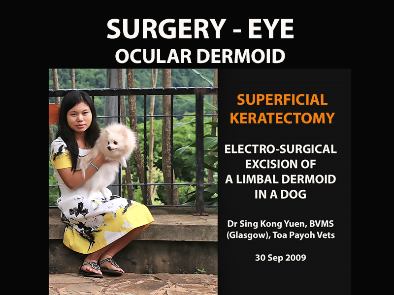
In this case, the video is about "How to excise a limbal dermoid using electrosurgery".
--------------------------------------
Shyan to re-make a new video, but note the following errors in the first video and use the amendments to create a really lively video.
1. INSERT THIS VIDEO HOOK WITH MY NARRATION
https://www.youtube.com/watch?v=I1gp0HgUVZE&feature=youtu.be
Use footage approx 00:0 - 1:04
2. Then continue with your present introduction...and continue
3. At Video 01:21. Insert TITLE. You narrated but did not insert the slide which would have been created by you. Use the following slide. It is important to show the title and narrate the text.

4. WHAT ARE OCULAR DERMOIDS AND WHAT TREATMENT WILL THE VET DO?
Dermoids are non-cancerous masses of fat, hair and skin found in an abnormal location of the body. Ocular dermoids are found in the eye. They are of two types.
Video 01:52. Replace the two dermoid slides with the following 3 slides and narrate them.


3. 2:05. Replace audio with the following:
Sedation is by IV ketamine and xylazine.
General anaesthesia is isoflurane gas and oxygen.
CONCLUSION.
TWO TYPES OF SURGICAL TREATMENT - SUPERFICIAL KERATECTOMY
3.1. Electro-surgery. The process is shown in this video.
3.2. Excision using surgical blade No. 11 and scissors.

The following shows
4. 2:57 Video footage of a Labrador Retriever puppy wearing an Elizabeth Collar after excision of the LIPODERMOID. The collar was won left for 14 days.
Narrate: "This image shows a Labrador Retriever wearing an Elizabeth Collar for 14 days. He had a LIPODERMOID excised using scalpel blade and scissors. "


Show the following and a video clip at:https://www.youtube.com/watch?v=EUq423Djdwg Use footage 0:00 - 0:09 around this clip section. Google how to download youtube video to get this video.
5.
03:12 Replace POST SUGERY with POST SURGERY.
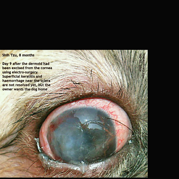
From the corneal healing at Day 9 when Dr Sing saw the dog, the outcome is excellent. No complaints from the owner for some months after the surgery as no news is good news.
6.
04:12 Create a proper attractive slide. For this video, use the following:

7. FINAL SLIDE. Should be "FOR MORE INFORMATION"....
use this slide: Narrate the text.

8. Good initiative. However, do not voice "thumbs up" as this is not in the script. Use video footage of Singapore scenes of HDB apartments and condos (instead of the two slides I gave you) using your smartphone to capture some clips if possible.
Intern to wait a day and view the video to catch such grammatical errors before submission to me.
---------------------------------------
THE FOLLOWING IS NOT PART OF THE SCRIPT. THEY ARE RESEARCH NOTES.
Dermoids are non-cancerous masses of fat, hair and skin found in an abnormal location of the body. Ocular dermoids are found in the eye. They are of two types. One is the LIPODERMOID . It is located in the conjunctival tissues near the lateral canthus. The other is the LIMBAL DERMOID. It is found in the limbus (junction of eye white and cornea). The surgical excision using electricity of this limbal dermoid forms the case study in this video. Shih Tzu puppies appear to be the top species to suffer from ocular dermoids according to cases seen at Toa Payoh Vets over the past 30 years. Surgical excision by Surgical blade No. 11 or in this case, by electro-excision are options of treatment. In this case, the owner did not want to leave his dog for 14 days as an inpatient. He took the dog back at Day 4. From the corneal healing at Day 9 when Dr Sing saw the dog, the outcome is excellent. No complaints from the owner for some months after the surgery as no news is good news.
-----------------------------
SUMMARY GIVEN IN YOUTUBE VIDEO
Dermoids are non-cancerous masses of fat, hair and skin found in an abnormal location of the body. Ocular dermoids are found in the eye. They are of two types. One is the LIPODERMOID . It is located in the conjunctival tissues near the lateral canthus. The other is the LIMBAL DERMOID. It is found in the limbus (junction of eye white and cornea).
The treatment is surgical excision using surgical blade No. 11 and scissors. I do not know whether other vets have had used electro-surgery as treatment.
The use of electricity to excise a limbal dermoid forms the case study in this video. Shih Tzu puppies appear to be the top species to suffer from ocular dermoids according to cases seen at Toa Payoh Vets over the past 30 years. Surgical excision by Surgical blade No. 11 or in this case, by electro-excision are options of treatment. In this case, the owner did not want to leave his dog for 14 days as an inpatient. He took the dog back at Day
----------------------------------------------
THIS IS PART OF THE NEW SCRIPT to be read together with the above mentioned amendments. Intern has to make a mind-map to connect all together in making a final script.
-----------------------------
15 Sep 2020. Script for Intern to create a BKTP video
(HOOK)
VIDEO HOOK WITH AUDIO
1. INSERT THIS VIDEO HOOK WITH MY NARRAION
https://www.youtube.com/watch?v=I1gp0HgUVZE&feature=youtu.be
Use footage approx 00:0 - 1:04
2. Then continue witH..................
Eyes are the first objects we see in a person or an animal. Pretty big clear eyes are attractive.

Eyes and a chrysanthemum-hair cut face make the Shih Tzu the top 3 small dog breeds as pets in Singapore.

From my Toa Payoh Vets case files over the past 40 years, the Shih Tzu breed has the most incidence of ocular dermoid.


Dermoids are non-cancerous masses with skin, hairs and fat. They can be found in people as well as dogs and cats.
There are two types. They are the limbal dermoid, being located in the limbus (sclera and corneal junction)
and the lipodermoid which is sited in the lateral canthal area (where the eyelids meet in the corner towards the ear).
As the hairs of the ocular dermoids cause eye irritation, most dogs feel uncomfortable and rubs the eyeball. The cornea becomes ulcerated and very painful.
What is the treatment?
Surgical excision of the dermoid. The procedure is known as superficial keratectomy

THIS IS A BE KIND TO PETS VETERINARY EDUCATIONAL VIDEO:

(Intern - to narrate the text in the slides above and below)
This video shows surgical procedures. Viewer discretion is advised.

SINGAPORE is a city state with more than 80% of the residents living in apartments. Small dog breeds such as the Shih Tzus are most popular as apartment pets.


The Shih Tzus appear to be the breed that is most affected by ocular dermoids, in cases seen at Toa Payoh Vets.


SUPERFICIAL KERATECTOMY - ELECTRO-SURGERY - (Narrate above slide)
I do not know whether other use electro-surgery to remove ocular dermoids from the cornea. This video shows the electro-surgical procedure to excise the limbal dermoid using electricity


A 3rd eyelid flap for 14 days facilitates healing of the exposed corneal epithelium. Antibiotic eye drops are applied daily for 14 days. An Elizabeth collar for 14 days prevents scratching of the eye. In young puppies and kittens of less than 2 months, anti-inflammatory eye drops must not be used to prevent swelling and scarring.

POST-SURGERY.
The owner did not permit me to ward the patient for 14 days after surgery. He wanted the dog back by Day 4. So, I took out the eyelid stitches sewing the eyelids together to facilitate corneal healing on Day 4 instead of Day 14.
On Day 9 after surgery, I followed up and saw the corneal healing taking place.
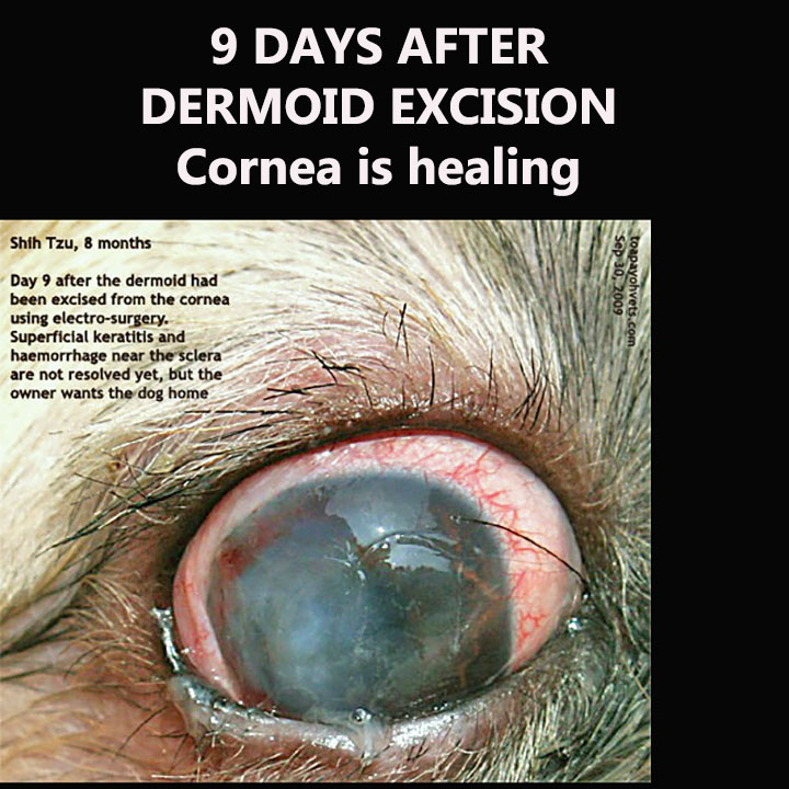
As to whether there will be corneal scarring later, I was unable to follow up. I electro-excised over 90% of the dermoid but not 100% as I did not want to risk the corneal laceration, rupturing the globe.
CONCLUSION.
SUPERFICIAL KERATECTOMY USING SURGICAL BLADE NO. 11 is the other option is to excise the limbal dermoid.
A stay suture to elevate the eyeball can be placed on the conjunctiva at the medial canthus area for easier surgery.
The outcome of the superficial keratectomy surgery is usually good. Care must be taken not to cut into the cornea in the excision of the limbal dermoid!
----------------------------------------------
22 Sep 2020.
THIS IS THE PREVIOUS SCRIPT - do not use this script.
15 Sep 2020. Script for Intern to create a BKTP video
(HOOK)
Eyes are the first objects we see in a person or an animal. Pretty big clear eyes are attractive.
Eyes and a chrysanthemum-hair cut face make the Shih Tzu the top 3 small dog breeds as pets in Singapore.

Dermoids are non-cancerous masses with skin, hairs and fat. They can be found in people as well as dogs and cats.
There are two types. They are the limbal dermoid, being located in the limbus (sclera and corneal junction)

and the lipodermoid which is sited in the temporal area (where the eyelids meet in the corner towards the ear - lateral canthus).

THIS IS A BE KIND TO PETS VETERINARY EDUCATIONAL VIDEO:
This video shows surgical procedures. Viewer discretion is advised.
SINGAPORE is a city state with more than 80% of the residents living in apartments. Small dog breeds such as the Shih Tzus are most popular as apartment pets.
The Shih Tzus appear to be the breed that is most affected by ocular dermoids, in cases seen at Toa Payoh Vets.
ELECTRO-SURGERY - EXCISION OF AN OCULAR DERMOID IN A DOG
Dr Sing Kong Yuen, BVMS (Glasgow), Toa Payoh Vets
I do not know whether other use electro-surgery to remove ocular dermoids from the cornea. This video shows the electro-surgical procedure to excise the limbal dermoid using electricity

SURGICAL PROCEDURES ARE AS FOLLOWS:
A 3rd eyelid flap for 14 days facilitates healing of the exposed corneal epithelium. Antibiotic eye drops are applied daily for 14 days. An Elizabeth collar for 14 days prevents scratching of the eye. In young puppies and kittens of less than 2 months, anti-inflammatory eye drops must not be used to prevent swelling and scarring.
POST-SURGERY.
The owner did not permit me to ward the patient for 14 days after surgery. He wanted the dog back by Day 4. So, I took out the eyelid stitches sewing the eyelids together to facilitate corneal healing on Day 4 instead of Day 14.
On Day 9 after surgery, I followed up and saw the corneal healing taking place.
As to whether there will be corneal scarring later, I was unable to follow up. I electro-excised over 90% of the dermoid but not 100% as I did not want to risk the corneal laceration, rupturing the globe.
CONCLUSION.
SUPERFICIAL KERATECTOMY USING SURGICAL BLADE NO. 11 is the other option is to excise the limbal dermoid.
A stay suture to elevate the eyeball can be placed on the conjunctiva at the medial canthus area for easier surgery.